The newborn infant’s head represents one-fourth of his total body length.
Its circumference is equal to that of his abdomen or chest. The average size is 13″ to 14″ (33-35 cm). The head is shaped or molded as it is forced through the birth canal in vertex presentations.
a. Molding.
During delivery, for the large head to pass through the small birth canal, the skull bones may actually overlap in a process referred to as molding. Such molding reduces the diameter of the skull temporarily. This elongated look usually disappears a few hours after birth as the bones assume their normal relationships (see figure 7-2).

b. Fontanels.
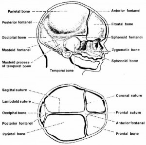
The infant’s skull is separated into six bones one from another along the suture lines (see figure 7-3). Where more than two bones come together, the space is called a fontanel. This is the unossified space or soft spot between the cranial bones of the skull in an infant. The infant’s pulse is sometimes visible there. The anterior fontanel is located at the intersection of the sutures of the two parietal bones and the frontal bones. It is diamond-shaped and strongly pulsatile. It normally closes at 9 to 18 months of age. The posterior fontanel is located at the junction of the sutures of the 2 parietal bones and 1 occipital bone. It is small, triangular shaped, and less pulsatile. It normally closes at 1 1/2 to 3 months of age. The anterior fontanel is the larger of the two.

c. Cephalohematoma.
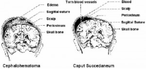
This is a collection of blood between a cranial bone and its overlying periosteum (see figure 7-4). Bleeding is limited to the surface of the particular bone. It is caused by pressure of the fetal head against the maternal pelvis during a prolonged or difficult labor. This pressure loosens the periosteum from the underlying bone, therefore rupturing capillaries and causing bleeding.
It may be apparent at birth but sometimes are not seen until 24 to 48 hours of life because subperiosteal bleeding is slow. It varies in size, rather firm to the touch and tends to increase in size from 1 to 3 days and then become softer and more fluctuant. Most cephalhematomas are absorbed within several weeks. No treatment is required in the absence of unexplained neurologic abnormalities.
d. Caput Succedaneum.
This is an abnormal collection of fluid under the scalp on top of the skull that may or may not cross the suture lines, depending on the size. Pressure on the presenting part of the fetal head against the cervix during labor may cause edema of the scalp (see figure 7-4). This diffuse swelling is temporary and will be absorbed within 2 or 3 days.

Brookside Note: The labels on Figure 7-4 above were apparently reversed in the original U.S. Army manual.
The drawing on the left actually illustrates a Caput Succedaneum, while the drawing on the right actually illustrates a Cephalohematoma.
