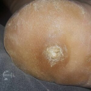
a. Common Warts (Verruca Vulgaris).
(1) Description. Warts of this type begin as smooth, flesh-colored papule. They may evolve into dome-shaped, gray-brown hyperkeratotic growths. Although these growths may be found on any skin surface, they most commonly occur on the hands.
(2) Treatment: keratolytic therapy. Different techniques are used to treat those warts. Keratolytic therapy and cryosurgery are two such techniques. See paragraph 1-7b(3) for a description of keratolytic treatment.

(3) Treatment: cryosurgery. This is treatment by liquid nitrogen and is performed in the following manner.
(a) Prepare a large cotton-tipped swab by winding the tip to a point.
(b) Dip the applicator into nitrogen.
(c) Immediately apply the tip to the center of the lesion.
(d) A white hard freeze will rapidly propagate in all directions.
(e) During the freezing process, the patient will experience pain that ranges from moderate to intense.
(f) Remove the swab after a 1 mm rim of freeze surrounding the lesion has been established.
NOTE: It is better to undertreat a benign lesion than to freeze too vigorously and destroy excessive amounts of normal tissue.
CAUTION: DO NOT use liquid nitrogen on a patient’s palms, soles, or areas that are automatically confined, such as the area around the nails. Swelling will occur in these confined areas.
b. Plantar Warts.
(1) Description. A plantar wart is a wart that occurs on the sole of the foot. Plantar warts occur at maximum pressure points; for example, over the heads of the metatarsal bones and on the heels. These warts are thick, painful calluses which have formed in response to pressure.
(2) Treatment: general. Treatment is not required as long as the warts are painless. It may be better not to subject the patient to a course of treatment but to let the wart go through the normal evolution. Severely painful plantar warts may be treated by keratolytic therapy (duofilm) or blunt dissection.
(3) Keratolytic therapy (duofilm). This type of treatment is conservative initial therapy. The treatment is nonscarring and relatively effective. It does require persistent application of medication once each day for several weeks. The procedure is given below.
(a) Pare down the wart with pumice stone or sandpaper.
(b) Soak the area in warm water to aid in the absorption of the medicine.
(c) Apply medicine with the glass rod and allow the medicine to dry.
(d) Cover the entire surface of the wart.
NOTE: Penetration of the medication is increased if the treated wart is covered with a piece of adhesive tape.
(e) After a few days, white, pliable keratin forms. Pare down this substance with sandpaper or a pumice stone.
(f) Eventually, you will expose pink skin.
(4) Blunt dissection. Perform the following steps.
(a) Inject 2% lidocaine with epinephrine directly into the substance of the wart.
(b) Insert the tip of a blunt-tipped scissors between the wart and normal skin.
(c) Cut the skin circumferentially.
(d) Insert a blunt dissector into the plane of cleavage.
(e) Separate the lesion with short firm strokes.
(f) Draw the blunt dissector firmly back and forth over the exposed surface of the bed to assure that no tissue fragments remain.
(g) Apply a small sterile dressing over the wound.
(h) Advise the patient to change the dressing daily for three to four days.
