
Two closely applied but separate membranes line the uterine cavity and surround the developing embryo-fetus.
Both membranes, the amnion (inner membrane) and the chorion (outer membrane), arise from the zygote. As the chorion develops, it blends with the fetal portion of the placenta; the amnion blends with the fetal umbilical cord.
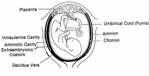
These deceptively strong, translucent membranes contain not only the fetus but also the amniotic fluid, and they are continuous with the margins of the placenta. See figure 2-7.
a. Amnion. This is the smooth, slippery, glistening innermost membrane that lines the amniotic space. It is filled with fluid and is often called the “bag of water.” The fetus floats and moves in the amniotic cavity. At full term, this cavity normally contains 500 cc to 1000 cc of fluid (water). This fluid provides many functions for the fetus. The amnion usually ruptures just before birth. The amnion functions to:
(1) Protect the fetus from direct trauma by distributing and equalizing any impact the mother may receive.
(2) Separate the fetus from the fetal membranes.
(3) Allow freedom of fetal movement and permits musculoskeletal development.
(4) Facilitate symmetric growth and development of the fetus.
(5) Protect the fetus from the loss of heat and maintains a relative, constant fetal body temperature.
(6) Serve as a source of oral fluid for the fetus.
(7) Act as an excretion and collection system.
b. Chorion. This is the outer membrane. It forms a large portion of the connective tissue thickness of the placenta on its fetal side. It is the structure in and through which the major branching umbilical vessels travel on the surface of the placenta.