These are the images used in this course. Click on any of the thumbnails to enlarge the image.
Figure 1.1 The Female Reproductive Organs. Sagittal Section
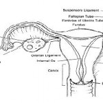
Figure 1-2. Anterior view of the uterus and related structures.
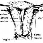
Figure 1-3. Walls of the uterus.
Figure 1-4. Human ovary.
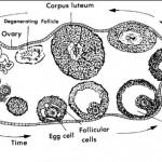
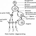
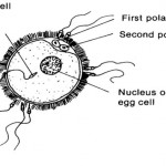
Figure 1-5. The process of oogenesis.
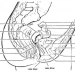
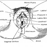
Figure 1-6. External female genitalia.
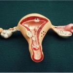
Uterus, Tubes, Ovaries and Vagina
Polycystic Ovary
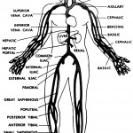
Vascular Tree
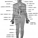
Arterial Vascular Tree
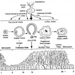
Figure 1-7. Menstrual cycle.
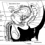
Figure 1-8. The male reproductive organs.
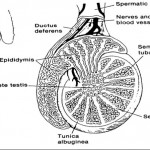
Figure 1-9. Structure of the testes.
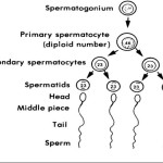
Figure 1-10. Spermatogenesis
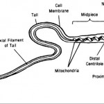
Figure 1-11. Structure of the sperm.
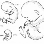
Figure 2.1 Growth of the Fetus
Figure 2-2. Sperm and ovum.
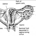
Figure 2-3. Travel of sperm to ovum.
Figure 2-4. Events of fertilization and implantation.
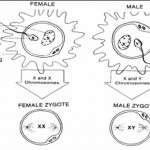
Figure 2-5. Genetic determination of sex.
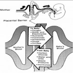
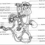
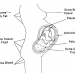
Figure 2-6. The placental circulation.
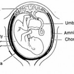
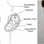
Figure 2-7. Fetal membranes.
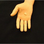
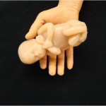
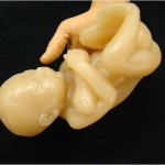
8 Week Fetus Model
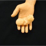
10 Week Fetus Model
15 Week Fetus Model
22 Week Fetus Model
32 Week Fetus Model
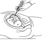
Figure 2-8. Amniocentesis.
Chromosome pattern of Down Syndrome (Trisomy 21) showing three “X” chromosomes instead of the usual two.
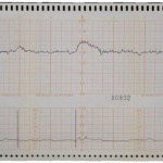
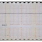
Illustration of a Reactive NST
Illustration of a Non-Reactive NST
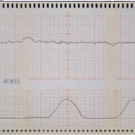
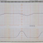
Illustration of a Negative OCT
Illustration of a Positive OCT
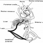
Figure 2-9. Fetal circulation before birth.
Figure 2-10. Fetal circulation after birth.
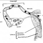
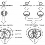
Figure 2-11. Development of twin fetuses.
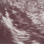
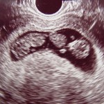
Ultrasound image of twins in the first trimester
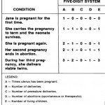
Table 3-1. Five-Digit System.
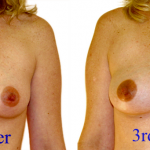
Breast Changes during Pregnancy
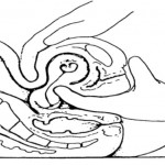
Figure 3.1 Hegars Sign
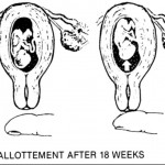
Figure 3.2 Ballotement
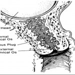
Figure 3.3 Cervix with Mucous Plug
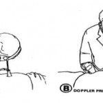
Figure 3-5. Detecting fetal heartbeat.
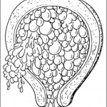
Figure 3.4 Hydatidiform Mole
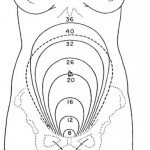
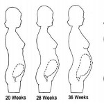
Figure 5-1. Appproximate height of the fundus at various weeks of pregnancy.
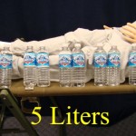
Total blood volume during pregnancy is about 5 liters
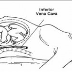
Figure 5-2. Vena cava syndrome.
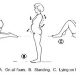
Figure 5-3. Postural changes during pregnancy.
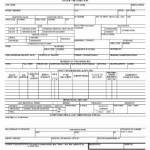
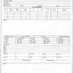
Figure 6-1A. BAMC Form 287 NS, Prenatal Questionnaire (front)
Figure 6-1B. BAMC Form 287 NS, prenatal Questionnaire (back).
Figure 6-2. SF 533, Medical Record–prenatal and Pregnancy
SF533
SF533 back
Figure 6-3. Patient in the lithotomy position, draped for pelvic exam.
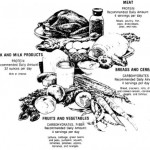
Figure 6-4. Four basic food groups.
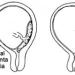
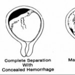
Figure 6-5. Various degrees of placenta previa.
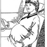
Figure 7-1. Seat belt wear.
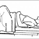
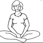
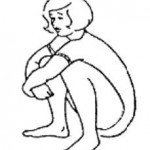
Figure 7-2. Tailor sitting exercise.
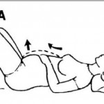
Figure 7-3. Pelvic rocking exercise.
Figure 7-4. Abdominal muscle contractions exercise.
Figure 7-5.Squatting exercise.
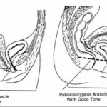
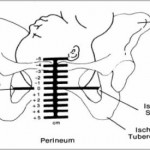
Figure 7-6. Muscles of the perineal area.
Figure 8-1. Relief of muscle cramp.
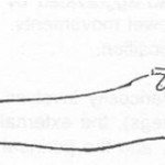
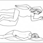
Figure 8-3. Positions for treatment of varicose veins.
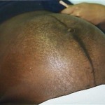
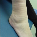
Ankle edema shown after her sock was removed
Figure 9-1. Distribution of normal weight gain.
Figure 9-1. Distribution of normal weight gain.
Figure 10-1. Typical types of presentations.
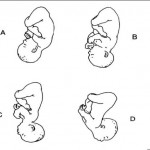
Figure 10-2. Types of attitudes. A–Complete flexion. B– Moderate flexion. C–Poor flexion. D–Hyperextension
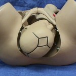
Figure 10-3. Measurement of station.
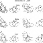
Figure 10-6. The mechanism of labor in the left occiput anterior (LOA) presentation.
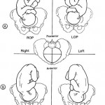
ROP (Right Occiput Posterior)
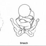
Figure 10-4. Breech positions.
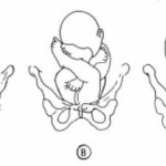
Figure 10-5. Examples of fetal vertex presentations in relation to quadrant of maternal pelvis.
Distance Learning for Medical and Nursing Professionals