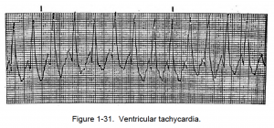
c. Ventricular Tachycardia. Analysis of ventricular tachycardia (VT) is given below and an example is shown in figure 1-31.
Ventricular tachycardia is serious and dangerous. It may be the precursor of ventricular fibrillation. If VT persists, there may be a marked reduction in cardiac output.
Lead_II_rhythm_ventricular_tachycardia_Vtach_VT. By Glenlarson (Own work) [Public domain], via Wikimedia Commons
(1) The rhythm of the heart is usually regular, but the rhythm can be slightly irregular.
(2) The heartbeat rate is from 150 to 250 beats per minute. The rate can exceed 250 beats per minute of the heart rhythm progressing to ventricular flutter. Occasionally, the rate may be slower than 150 beats per minute; the condition is then termed slow ventricular tachycardia (VT).
(3) P waves are not normally seen; however, dissociated waves may be seen. The focus is in the ventricles, and there will be no PR interval.
(4) The QRS complex is wide and bizarre. The complex may be 0.12 of a second or greater.
(5) The T wave is usually in the opposite direction from the R wave.


![1-11. VENTRICULAR ARRHYTHMIAS: C. Ventricular Tachycardia 1 Lead_II_rhythm_ventricular_tachycardia_Vtach_VT. By Glenlarson (Own work) [Public domain], via Wikimedia Commons](https://brooksidepress.org/ecg/wp-content/uploads/2015/11/Lead_II_rhythm_ventricular_tachycardia_Vtach_VT-300x58.jpg)