a. General Information. Heart blocks are arrhythmias caused by conduction disturbances at the AV node.
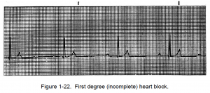
b. First-Degree (Incomplete) Heart Block. The first-degree heart block takes place where there is an incomplete block. Analysis of first degree heart block is given below and an example is shown in figure 1-22.
First Degree Heart Block. By Michael Rosengarten BEng, MD.McGill [CC BY-SA 3.0 (http://creativecommons.org/licenses/by-sa/3.0)], via Wikimedia Commons
(1) The regularity and rate depend on the underlying rhythm of the heart.
(2) The P waves are upright and uniform and followed by the QRS complex.
(3) The PR interval is constant and greater than 0.20 seconds.
(4) The QRS complex is less than 0.12 seconds.

c. Wenckebach (Second-Degree Heart Block/Mobitz Type I). This condition is a less serious type of second-degree heart block.
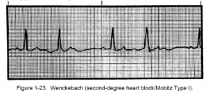
It is, however, still quite common in patients with acute myocardial infarction. The condition may be produced by digitalis. Analysis of second degree heart block (Mobitz Type I) is given below and an example is shown in figure 1-23.
![1-10. HEART BLOCKS 3 Second-Degree Heart Block:Mobitz Type I. By James Heilman, MD (Own work) [CC BY-SA 3.0 (http://creativecommons.org/licenses/by-sa/3.0)], via Wikimedia Commons](https://brooksidepress.org/ecg/wp-content/uploads/2015/11/Second-Degree-Heart-BlockMobitz-Type-I-300x32.png)
(1) The rhythm is irregular, and the R-R interval gets progressively shorter as the P-R intervals get longer.
(2) Grouped beating is characteristic.
(3) The heartbeat rate is usually slightly slower than normal.
(4) P waves are upright and uniform, but some P waves are not followed by QRS complexes.
(5) The P-R interval progressively lengthens until one P wave is not conducted.
(6) The QRS complex lasts less than 0.12 of a second.

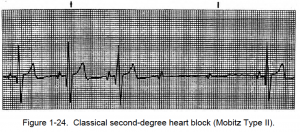
d. Classical Second-Degree Heart Block (Mobitz Type II). Analysis of second degree heart block (Mobitz Type II) is given below and an example is shown in figure 1-23.
![1-10. HEART BLOCKS 5 Second degree heart block type 2. By James Heilman, MD (Own work) [CC BY-SA 3.0 (http://creativecommons.org/licenses/by-sa/3.0)], via Wikimedia Commons](https://brooksidepress.org/ecg/wp-content/uploads/2015/11/Second-degree-heart-block-type-2-300x30.png)
(1) The rhythm is regular if the conduction ratio is constant; however, the rhythm is irregular if the conduction ratio varies.
(2) The rate for atrial is usually normal and the ventricular rate is usually bradycardia.
(3) P waves are upright and uniform with more P waves than QRS complexes (usually a ratio of 2:1, 3:1, and so forth.).
(4) The P-R interval is constant on conducted beats, but it may be longer than normal.

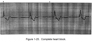
e. Complete Heart Block (Third-Degree). Analysis of third degree heart block is given below and an example is shown in figure 1-25.
![1-10. HEART BLOCKS 7 Complete Heart Block (Third-Degree). By James Heilman, MD (Own work) [CC BY-SA 3.0 (http://creativecommons.org/licenses/by-sa/3.0)], via Wikimedia Commons](https://brooksidepress.org/ecg/wp-content/uploads/2015/11/Complete-Heart-Block-Third-Degree-300x22.png)
(1) The rhythm is regular with PP and RR intervals constant.
(2) The atrial rate is normal, but the ventrical response rate varies in this way. The junction focus has a rate of 40 to 60 beats per minute. The ventricle focus has a rate of 20 to 40 beats per minute.
(3) P waves are upright and uniform with more P waves than QRS complexes.
(4) There are no PR intervals because the P waves have no relationship to QRS complexes. Occasionally, a P wave is superimposed on a QRS complex.
(5) The QRS complex is less than 0.12 seconds at the junctional focus and greater than 0.12 seconds at the ventricular focus.
(6) Cardiac output may be greatly diminished if the heart rate is below 35 to 50 beats per minute. Additionally, in third-degree heart block, the atria and ventricles are no longer synchronized; therefore, the ventricles do not fill completely before each contraction, causing cardiac output to be even further reduced. The ventricular rate may be so slow that circulation cannot be maintained and syncope (congestive heart failure) or angina may occur.

