a. Regularity.
(1) Determine regularity (also called rhythm) by looking at the R to R interval (RRI). This interval can be regular or irregular.
(2) The R-R interval is constant for regular rhythm, which means the distance between the beats, is the same. See figure 1-7 for an example of regular rhythm.
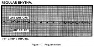
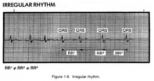
(3) If the R to R interval (RRI) is inconsistent and the rhythm is irregular, the heartbeats are premature. A basically regular rhythm occurs when a heartbeat or two interrupt a regular rhythm; for example, ectopic (heartbeats originating from an abnormal place; that is, from somewhere other than the S-A node) or grouped heartbeats. A regularly irregular rhythm is a pattern of irregularity.
A totally irregular rhythm has no pattern at all. See figure 1-8 for an example of irregular rhythm.
b. Rate.
(1) Rate for regular heartbeat rhythms can be calculated in two ways.
(a) Method I. The most accurate method is to count the number of small squares between the two R waves and divide that number into 1500.
(b) Method II. The faster method is to count the number of large squares between R waves and divide that number into 300. This method is useful during an emergency. This method does not require a 6-second strip. Also, it allows rapid rate calculation using simple memorization of rate for large boxes.
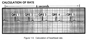
(2) The heartbeat rate for irregular heartbeat rhythm is based on the 6-second strip method. The minimum amount of time needed to determine cardiac rhythm is 6 seconds. The method described here is the quickest and easiest method although it is not as accurate as the “box” method. The vertical notches in the upper margin of the paper are 3 seconds apart.
(a) Count the number of QRS complexes in a 6-second strip. See figure 1-9.
(b) Multiply by 10 to get the heart rate for one minute.
c. P Waves. P waves are small positive deflections that occur on the oscilloscope.
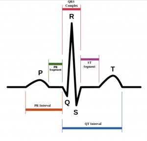
These waves represent atrial depolarization. Normally, the P waves are rounded, uniform, and upright and come before the QRS complex. These waves are either regular or irregular. Measured distances between all P waves should be constant. Determine whether there is a P wave for every QRS complex. Determine whether the P wave is in front of the QRS complex and see if there are more P waves than QRS complexes. Check to see if all the P waves look alike. See if there are irregular P waves associated with ectopic or irregular beats.
d. P-R Intervals. All P-R intervals should be constant.
Measure the P-R intervals to determine if the intervals are normal or abnormal. A normal P-R interval is from 0.12 to 0.20 seconds. If the P-R interval varies, notice if there is a pattern to the changing measurements.
e. QRS Complexes. These complexes represent ventricular depolarization. The QRS complexes should be of equal duration.
(1) An QRS example is shown in figure 1-10. An analysis of the parts of the QRS complex is given below.
(a) Q waves are a negative deflection on the oscilloscope. They follow the P waves.
(b) R waves are a positive deflection on the oscilloscope, and they follow the Q waves.
(c) S waves are a negative deflection on the oscilloscope following the R waves.
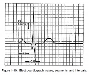
Figure 1-10. Electrocardiograph waves, segments, and intervals.
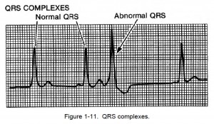
(2) Check the measurement of the QRS complex. The complex should be less than 0.12 seconds. See if the measurement is within normal limits. See if all the QRS complexes look alike. If there are any unusual QRS complexes, determine if they are associated with ectopic beats (abnormal heartbeats). Figure 1-11 shows an example of an abnormal QRS.
NOTE: For instructional purposes, the lines on the figures showing heartbeats are sometimes heavier than normal.
