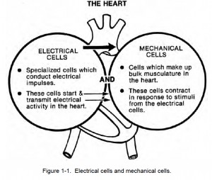
a. Heart Cells. Two types of cells are found in the heart — electrical cells and mechanical cells (see figure 1-1).
The heart’s conduction system is made up of electrical cells. These cells have the ability to begin and transmit electrical activity in the heart.
Myocardial cells (the mechanical cells) make up the bulk musculature of the heart. When an electrical stimuli reaches these cells, the cells contract. An electrical impulse stimulates the mechanical action of the heart causing the heart to pump effectively. If the electrical system of the heart does not function properly, arrhythmias (a mechanical activity) may occur.

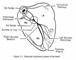
b. Electrical Activity In The Heart. The heart’s electrical activity begins in the sinoatrial (SA) node and flows toward the ventricles (see figure 1-2).
The SA node is the heart’s pacemaker. All the areas of this conduction system initiate impulses, become irritable, and respond to an impulse. Impulses are initiated in each area of the conduction system as shown below.
SA Node………..60-100 per minute
AV Junction……40-60 per minute
Ventricle ………..20-40 per minute

c. The Pacemaker Site. The common pacemaker site is the SA node because it initiates electrical impulses at a faster rate than the junction or ventricle.
An impulse from the AV junction can take over if the SA node should fail. If the AV junction also fails, the ventricle can take over. This is a protective backup system, a system which
helps the heart maintain electrical efficiency. Sometimes the junction or the ventricle becomes irritable and starts impulses at a faster than normal rate which overrides the SA node. When this happens, the pacemaker site that is the fastest dominates and takes over control of the heart.
