a. Normal Sinus Rhythm (NSR).
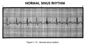
(1) Normal sinus rhythm originates in the SA node (pacemaker) and travels through the normal conduction pathways. It is not an arrhythmia (abnormality in the normal cardiac rhythm) or dysrhythmia (a disturbance in cardiac rhythm) because it is a normal pattern. See figure 1-12.
(2) Normal sinus rhythm is analyzed in this manner:
(a) When the R-R intervals and the P-P intervals are constant, the rhythm is considered regular.
(b) The atrial and ventricular rates equal 60 to 100 heartbeats per minute with no added or lost P, QRS, or T waves.
(c) The P wave has a uniform configuration with one P wave in front of every QRS.
(d) The P-R interval is constant between 0.12 and 0.20 seconds.
(e) The QRS complex measures less than 0.12 seconds.

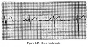
b. Sinus Bradycardia. The analysis of sinus bradycardia is given below and an example shown in figure 1-13.
![1-07. CARDIAC RHYTHMS 2 Sinus Bradycardia. By Sinusbradylead2.JPG: James Heilman, MD derivative work: Mysid (using Perl and Inkscape) (This file was derived from Sinusbradylead2.JPG:) [CC BY-SA 3.0 (http://creativecommons.org/licenses/by-sa/3.0) or GFDL (http://www.gnu.org/copyleft/fdl.html)], via Wikimedia Commons](https://brooksidepress.org/ecg/wp-content/uploads/2015/11/800px-Sinus_bradycardia_lead2.svg_-300x102.png)
(1) The rhythm is regular with the R-R intervals constant and the P-P intervals constant.
(2) The atrial and ventricular rates are less than 60 beats per minute.
(3) The P wave is normal and upright with one P wave in front of every QRS complex.
(4) The P-R interval is constant between 0.12 and 0.20 seconds.
(5) The QRS complex measures less than 0.12 seconds.
(6) A heart rate of less than 60 beats per minute may indicate good physical conditioning if the individual is young and healthy.
(7) If the person is suffering from acute myocardial infarction (AMI), this heartbeat rate may indicate any one of the following: conduction system damage, increased parasympathetic tone, or possible toxic levels of certain cardiac drugs (digitalis, quinidine).
If the heartbeat rate decreases to less than 50 beats per minute, the heart may not be able to pump enough fluid through the body’s vital organs. Additionally, bradycardia leads to electrical instability in the ventricles, possibly resulting in ventricular arrhythmias.
NOTE: Males normally have a slower heart rate than females. Cardiovascularly healthy people may normally have a slow (less than 60 beats per minute) heart rate which also normally slows during sleep and rest. A slow heart rate is significant only if it is associated with MI or cardiovascular compromise.

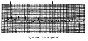
c. Sinus Tachycardia. The analysis of sinus tachycardia is given below and an example is shown in figure 1-14.
![1-07. CARDIAC RHYTHMS 4 Sinus Tachycardia. By James Heilman, MD (Own work) [CC BY-SA 3.0 (http://creativecommons.org/licenses/by-sa/3.0)], via Wikimedia Commons](https://brooksidepress.org/ecg/wp-content/uploads/2015/11/sinus_tachycardia-300x96.png)
(1) In sinus tachycardia, the rhythm is regular and the R-R and P-P intervals are constant.
(2) Atrial and ventricular rates are equal to or greater than 100 beats per minute.
(3) The P wave is normal and upright with one P wave in front of every QRS complex.
(4) The P-R interval is constant between 0.12 and 0.20 seconds.
(5) The QRS complex measures less than 0.12 seconds.
(6) A variety of circumstances can cause sinus tachycardia including pain, fever, hypoxia, shock, congestive heart failure, and drugs such as epinephrine, atropine, and isoproterenol. The more rapid the heart rate, the harder the heart works. This can lead to further heart damage in AMI.
Also, because there is insufficient time between contractions for the ventricles to fill completely with blood, the heart may not be able to pump fluid effectively when the heart rate is more than 120 to 140 beats per minute. Strenuous exercise such as jogging may cause this condition.

