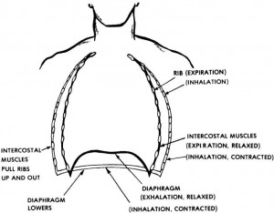
Ventilation is caused by two muscle systems–the diaphragm and the intercostal muscles.
When the diaphragm and the intercostal muscles contract (get shorter), they make the chest cavity larger. The lungs then expand in order to fill up the space. When the lungs expand, air from the outside environment rushes in through the mouth or nose to fill up this extra space.
When the muscles relax, the chest cavity returns to its normal size. This action compresses the air in the lungs and forces in some of the air from the lungs, through the windpipe, and out of the nose or mouth.
a. Diaphragm.
The diaphragm is a large dome-shaped muscle that separates the chest cavity from the abdominal cavity. When the diaphragm contracts, the muscle flattens somewhat and “lowers the floor” of the chest cavity (figure 4-1).
When the muscle relaxes, it returns to its normal (dome) shape. The diaphragm is responsible for most of the air movement during breathing. The diaphragm is a skeletal muscle that is under involuntary control of the part of the brain that controls breathing.
b. Intercostal Muscles.
The intercostal muscles are the muscles that connect one rib to another rib. When the muscles contract (shorten), the ribs are pulled up and out. This action causes the entire rib cage to move up and out (away from the body) as illustrated in figure 4-1. This up and out motion causes the circumference of the chest to increase.

