Skip to content
Figure 8-8. Leg exercises.
Figure 8-7. Closed suction device.
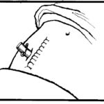
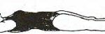
Figure 8-6. Penrose drain.
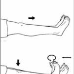
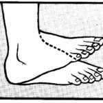
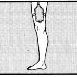
Figure 8-5. Dorsiflection of the foot.
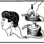
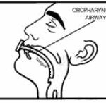
Figure 8-4. Oropharyngeal airway.
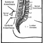
Figure 8-3. Sites for spinal anesthetics
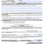
Figure 8.2. SF 522, Request for Administration of Anesthesia and for Performance of Operations and Other Procedures.
Figure 8-1. DD Form, 1924, Surgical Check List.
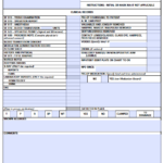
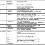
Table 7-1. Selected teaching strategies.
Figure 7-1. Example of a contractual agreement.
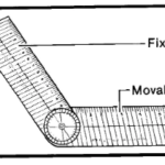
Figure 6-9. Goniometer.
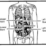
Figure 6-8. Abdominal regions.
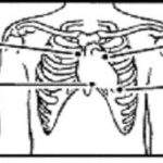
Figure 6-7. Areas to auscultate for heart sounds.
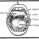
Figure 6-6. Oral cavity.
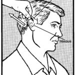
Figure 6-5. Movement of the auricle.
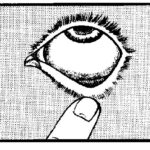
Figure 6-4. Exposing sclera and conjuctiva.
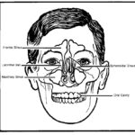
Figure 6-3. Anatomy of the skull.
Figure 6-2. Percussion.
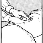
Palpation of the abdomen
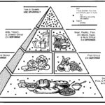
Figure 5-1. Food Guide Pyramid.
Example
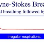
Cheyne Stokes Breathing
Slow Breathing
Tachypnea – Rapid Breathing
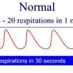
Normal Breathing
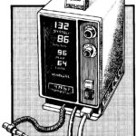
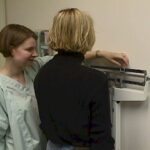
Figure 4-3. Electronic vital signs monitor.
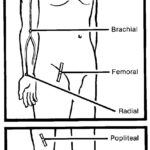
Figure 4 –2. Arterial pulse sites.
Figure 4-1. Clinical thermometers.
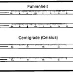
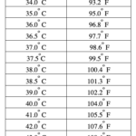
Table 4-2. Celsius/Fahrenheit equivalent temperature.
Weigh the patient in the same amount of clothing each day.
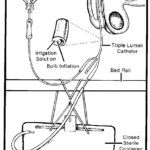
Figure 3-9. Foley triple lumen catheter.
Figure 3-8. Urinary collecting leg bag.
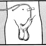
Figure 3-7. Securing (female) indwelling catheter.
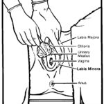
Figure 3-6. Inserting the catheter in a female.
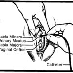
Figure 3-5. Cleansing the female meatus.
Figure 3-4. Anchoring (male) indwelling catheter.
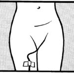
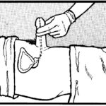
Figure 3-3. Positioning the penis at a 90-degree angle
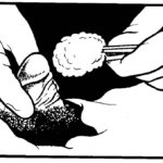
Figure 3-2. Cleansing the male meatus.
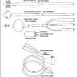
Figure 3-1. Urinary catheters.
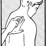
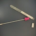
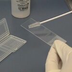
Sterile cotton-tipped applicator specimen collection kit (culturette).
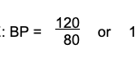
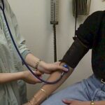
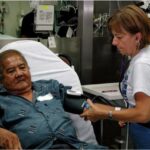
Take the blood pressure 10-15 minutes after the patient has rested.
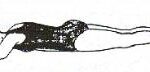
Figure 1-7. Knee-chest position.
Figure 1-6. Sim’s position.
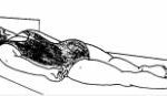
Figure 1-5. Prone position.
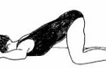
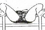
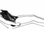
Figure 1-4. Dorsal lithotomy position.
Figure 1-3. Fowler’s position.
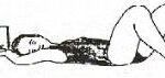
Figure 1-2. Dorsal recumbent position.
Figure 1-1. Horizontal recumbent position.
Basic Nursing Education