Cardiopulmonary Resuscitation
LESSON 1: REVIEW OF THE CIRCULATORY AND RESPIRATORY SYSTEMS
1-4
1-4. BLOOD FLOW
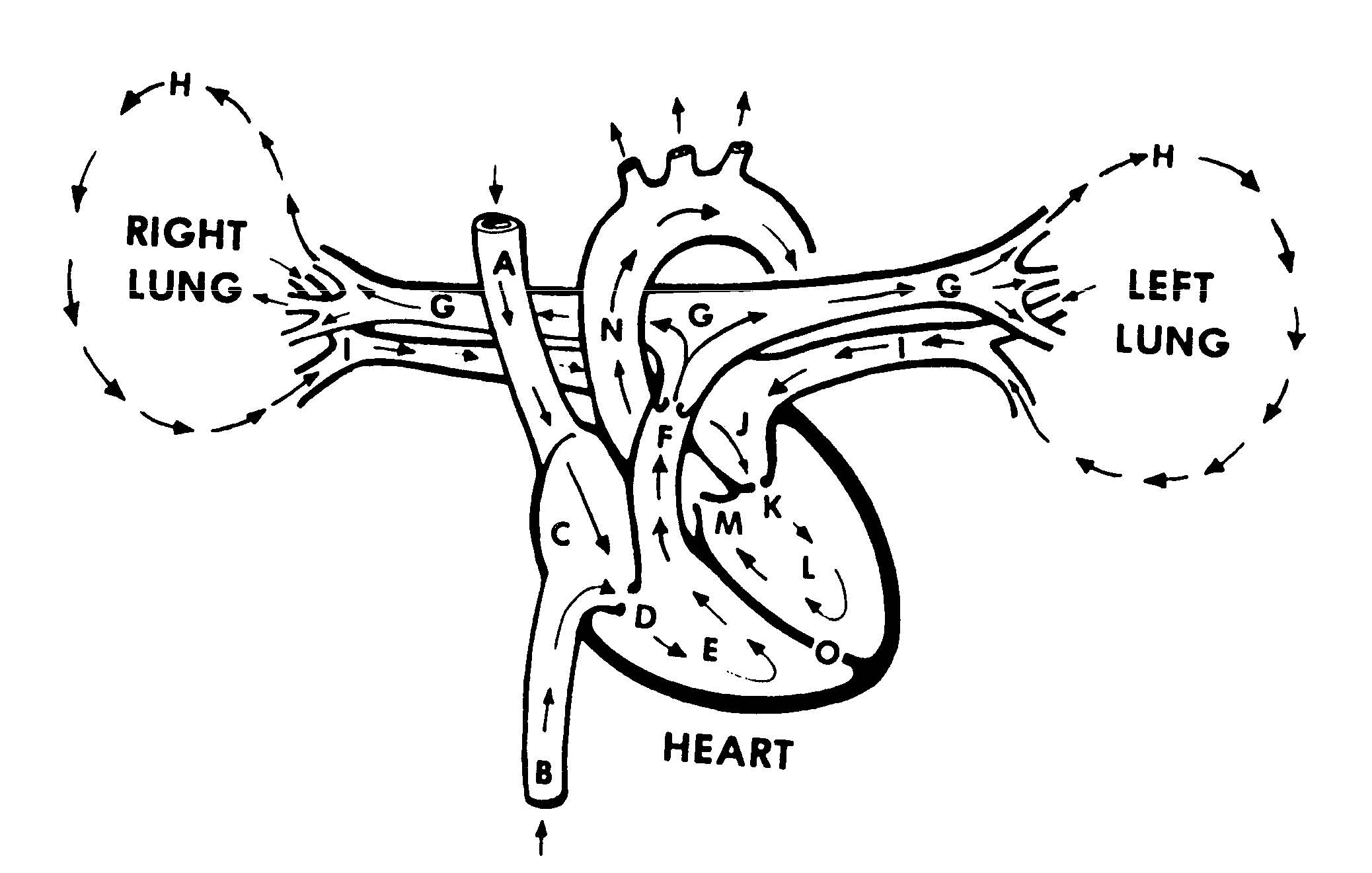
In order to summarize how blood flows in the body, let's take a trip through the body's circulatory system (figure 1-2). We will enter the system at the vena cava.
a. Vena Cava. There are two major blood veins, which empty into the right atrium. The superior vena cava carries oxygen-poor blood coming from the head, arms, and chest. The inferior vena cava returns oxygen-poor blood from the lower trunk and legs.
|
A. Superior vena cava I. Pulmonary veins |
|
B. Inferior vena cava J. Left atrium |
|
C. Right atrium K. Mitral valve |
|
D. Tricuspid valve L. Left ventricle |
|
E. Right ventricle M. Aortic valve |
|
F. Pulmonary valve N. Aorta |
|
G. Pulmonary arteries O. Interventricular septum |
|
H. Lungs |
Figure 1-2. Blood flow to and from the heart (not drawn to scale, front view).
b. Right Atrium. The right atrium receives blood from the superior vena cava and the inferior vena cava. When the right ventricle relaxes (that is, after it has contracted and pumped blood), blood flows from the right atrium into the right ventricle through the tricuspid valve. The tricuspid valve is formed so that blood cannot flow back into the right atrium when the right ventricle contracts.
c. Right Ventricle. When the right ventricle is filled with blood, it receives an impulse from the sinoatrial node. This impulse causes the muscles of the right ventricle to contract. This contraction causes the inside of the ventricle (the space where the blood is) to become smaller. The increased pressure forces blood out of the ventricle and into the pulmonary artery. The pulmonary valve located at the beginning of the pulmonary artery keeps blood from flowing back into the right ventricle when the ventricle relaxes and returns to its normal size.
d. Lungs (Pulmonary System). The pulmonary artery divides into two arteries. One artery travels to the right lung while the other artery travels to the left lung. The arteries divide until they reach the capillary stage. The capillaries surround the alveoli (air sacs) of the lungs. There the oxygen-poor blood gets rid of carbon dioxide and picks up oxygen from the air in the alveolus. The blood, now high in oxygen content, then returns to the left atrium through the pulmonary veins.
e. Left Atrium. The left atrium receives blood from the lungs through two pulmonary veins. When the left ventricle relaxes after having contracted, the blood flows from the left atrium into the left ventricle through the mitral valve. The mitral valve keeps blood from flowing back into the left atrium when the left ventricle contracts.
f. Left Ventricle. After the left ventricle is filled with oxygen-rich blood, it receives an impulse from the sinoatrial node, which causes it to contract and pump blood into the large artery call the aorta. When the blood enters the aorta, it passes through the aortic valve. This valve keeps the blood from flowing back into the heart once the left ventricle relaxes.
g. Body (Systemic System).
(1) Some arteries branch off the aorta to provide the brain, upper body, and heart with blood. Blood returns to the heart from these areas through the superior vena cava.
NOTE: If blood flow to the brain stops and is not restored (either the casualty's heart
starts beating on its own or cardiopulmonary resuscitation is administered),
the brain will begin to die in six to ten minutes.
(2) The aorta turns down and divides into smaller arteries which go to the lower parts of the body. Some of the blood picks up fluids and nutrients from the intestines. Some of the blood passes through the liver and kidneys which remove bacteria and other unwanted substances from the blood. The blood returns to the heart from these areas through the inferior vena cava.