Cardiopulmonary Resuscitation
LESSON 1: REVIEW OF THE CIRCULATORY AND RESPIRATORY SYSTEMS
1-5
1-5. THE RESPIRATORY SYSTEM
The respiratory system consists of two lungs and the respiratory tract that carries air to and from the lungs (figure 1-3). When a person inhales, the air enters the nose or mouth, travels down the trachea, and into the two bronchi. Each bronchus divides into smaller and smaller air tubes. Finally, the air reaches the alveoli (air sacs). The red blood cells in the capillaries surrounding the alveoli absorb oxygen from the air and give off carbon dioxide, which passes into the alveoli. When a person exhales, the air travels from the alveoli through the air tubes, up the trachea, and out the nose or mouth. Of course, not all of the air inhaled reaches the alveoli nor is all of the oxygen removed from the air in the alveoli. The average adult takes in about 500 milliliters (ml) of air each time he inhales and he exhales the same amount. Even after the person exhales, the lungs still contain about 2300 ml of air. The anatomy (structures) and the physiology (functions) of the respiratory system are briefly discussed below.
a. Nose. The nose is composed of two nostrils (openings) and two nasal cavities (air chambers above the roof of the mouth and below the cranium). A structure called the nasal septum separates the right nostril and nasal cavity from the left nostril and nasal cavity. The nose warms, moistens, and filters the inhaled air. Special nerve endings in the upper part of the nasal cavities provide the sense of smell.
b. Pharynx. The pharynx is a part of the throat that is part of both the respiratory system and the digestive system. The pharynx is divided into three parts. The nasopharynx (upper part) connects with the nasal chambers. The oropharynx (middle part) connects with the oral cavity (mouth). The laryngopharynx (lower part) connects with the larynx (respiratory system) and the esophagus (digestive system).
c. Epiglottis. The epiglottis is a flap that covers the entrance to the larynx when a person swallows. This prevents food from entering the larynx instead of the esophagus. When a person inhales, the entrance to the larynx is not covered and air enters the larynx. If a foreign object enters the airway, it can block the airway and cause breathing to stop.
d. Larynx. The larynx is a box-like structure composed of cartilage, ligaments, and muscles that sits on top of the trachea. The larynx contains the vocal cords, which produce the voice; therefore, it is sometimes called the voice box. It is also called the Adam's apple because of the bulge it causes in the throat.
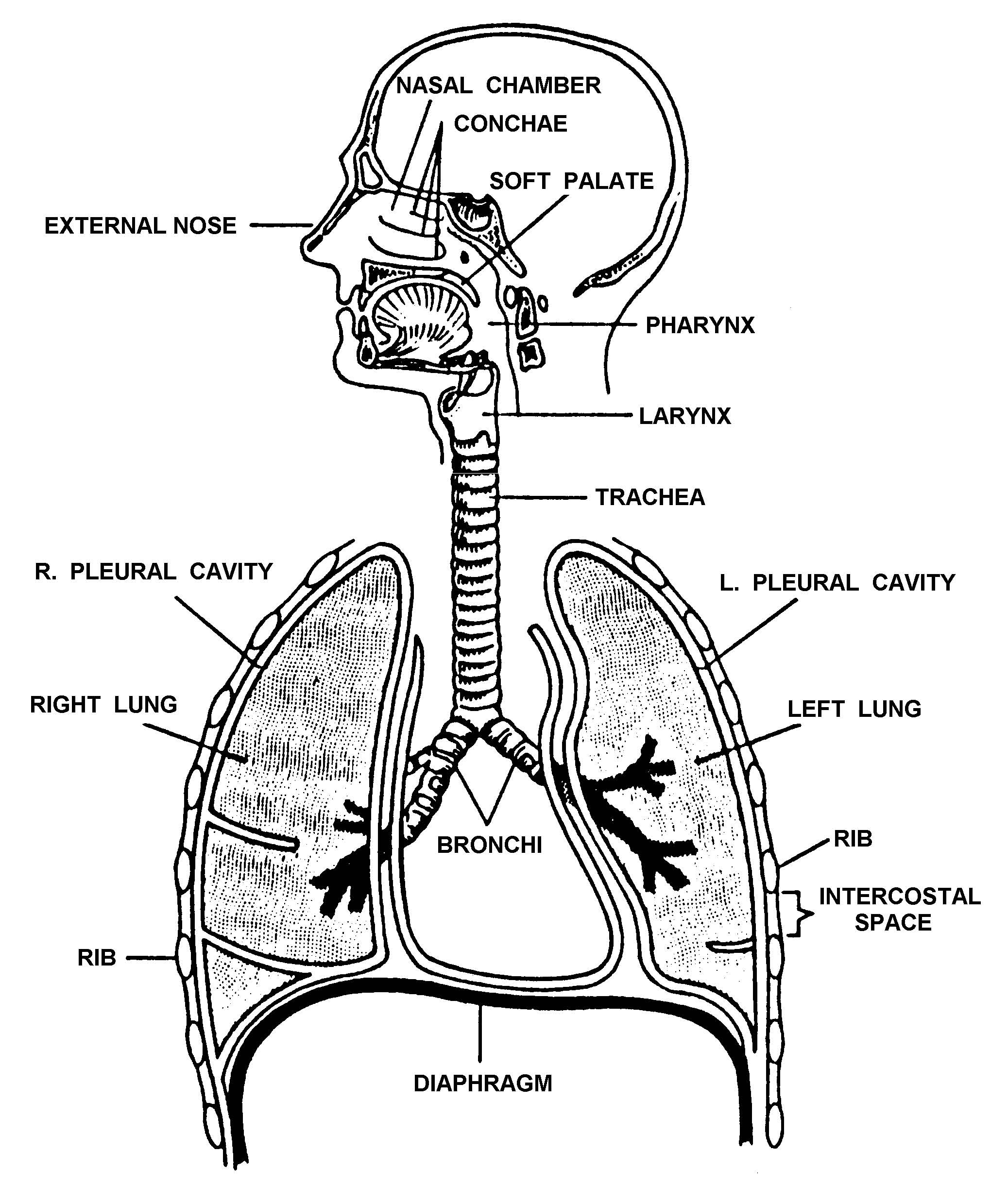
Figure 1-3. The respiratory system.
e. Trachea. The trachea (windpipe) is a tube composed of horseshoe-shaped rings of cartilage. Cilia (hair-like projections) on the inner lining of the trachea help to filter air as it passes through the trachea to the bronchi.
f. Bronchi. The bronchi are two tube-like structures at the base of the trachea. One bronchus leads toward the right lung; the other bronchus leads toward the left lung. Like the trachea, the bronchi are composed of cartilaginous rings and are lined with a mucous membrane.
g. Bronchioli. The bronchi divide and subdivide until they become small air tubes one millimeter or less in diameter call bronchioli. The bronchioli continue to subdivide until they become very small tubes ending in alveoli.
h. Alveoli. Alveoli are tiny, grape-like clusters of microscopic air sacs. Air enters the alveoli from the bronchioli. The wall of an alveolus is one cell layer thick. The alveolus is surrounded by equally thin
capillaries. Oxygen (O2) molecules from the air inside the alveolus travel through the alveolus and capillary walls to the blood within the capillary. The hemoglobin in the red blood cells captures the oxygen molecules and release carbon dioxide (CO2) molecules. The carbon dioxide molecules and some water molecules travel from the blood, through the walls, and into the alveoli.
i. Lungs. The alveoli, bronchioli, and associated blood vessels make up two cone-like organs called lungs. The lungs are broad at their base (which rests on the diaphragm) and narrow at the apex (top). Each lung is surrounded by pleural membranes that prevent friction when the lung expands and contracts. The right lung is divided into three lobes; the left lung is divided into two lobes. The left lung is smaller than the right lung because the heart takes up space on the left side of the chest cavity.