Cardiopulmonary Resuscitation
LESSON 1: REVIEW OF THE CIRCULATORY AND RESPIRATORY SYSTEMS
1-3
1-3. THE CIRCULATORY SYSTEM
The circulatory system consists of the heart, blood vessels, and blood. The circulatory system brings oxygen and nutrients to the body's cells and carries away waste products. The circulatory system is also called the cardiovascular system ("cardio-" means heart; "-vascular" means vessels.)
a. Heart. The heart (figure 1-1) is a strong, muscular organ that, by its rhythmic contractions, acts as a force pump maintaining blood circulation. The heart is about the size of a fist and is located in the lower left-central part of the chest cavity.
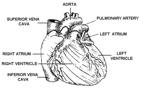
Figure 1-1. The human heart (front view).
(1) Layers. The heart consists of three layers.
(a) The myocardium is the middle layer. It is composed of the actual heart muscles. ("Myo-" means muscle; "cardium" means heart.)
(b) The pericardium is the outer layer. It is a double-walled sac that surrounds the heart muscles. ("Peri-" means around.)
(c) The endocardium is the inner layer. It forms the inner lining of the four chambers. ("Endo-" means within.)
(2) Chambers. The heart can be described as being two pumps. Each side (right half and left half) of the heart has a receiving chamber for the blood (the atrium) and a pumping chamber (the ventricle). The two halves of the heart are separated by a wall-like structure called the interventricular septum.
NOTE: The plural of atrium is atria.
(3) Sinoatrial node. The sinoatrial (SA) node is a small bundle of nerve tissue located at the junction of the superior vena cava and the right atrium. The sinoatrial node is a natural pacemaker that produces an electrical stimulus. This electrical stimulus causes the muscles of the ventricles to contract and pump blood.
b. Blood Vessels. The blood vessels are firm, elastic, muscular tubes that carry the blood away from the heart and back to the heart again.
(1) Blood circulation systems. Since the heart is divided into two parts (the right half consisting of the right atrium and the right ventricle and the left half consisting of the left atrium and left ventricle), it is not surprising to find that there are actually two blood circulatory systems--the systemic and the pulmonary.
(a) Systemic. The systemic (general) circulatory system is the larger of the two systems. It takes the blood pumped by the left ventricle to all parts of the body and returns the blood to the right atrium. The oxygen content of the blood is high when it leaves the heart through the left ventricle and is low when it returns to the right atrium.
(b) Pulmonary. The pulmonary circulatory system takes the blood pumped by the right ventricle to the lungs and returns the blood to the left atrium. The oxygen content of the blood is low when it leaves the heart through the right ventricle and high when it returns to the left atrium.
(2) Types of blood vessels. Both the systemic and the pulmonary circulatory systems are composed of three major types of blood vessels--arteries, capillaries, and veins.
(a) Arteries. The arteries carry blood pumped by the ventricles away from the heart. The arteries of the systemic circulatory system carry oxygenated (oxygen rich) blood to body tissues. The pulmonary arteries carry deoxygenated (oxygen-poor) blood to the lungs. Arteries have the capacity to constrict and dilate. This constricting and dilating helps to regulate the blood pressure.
(b) Capillaries. Originally, the arteries are large blood vessels. Soon, however, they divide into smaller branches. These branches then divide again and again. With each division, the blood vessels become smaller and smaller. Finally, the blood vessels are so small that only one red blood cell can pass through at a time. When they reach this size, the blood vessels are called capillaries. When a
red blood cell enters the capillaries, it is free to perform its primary functions.
In the pulmonary system, red blood cells give up carbon dioxide to the lungs and pick up oxygen. In the systemic system, red blood cells give oxygen and nutrients to the cells and pick up carbon dioxide and other waste products.
(c) Veins. Capillaries join together to form larger blood vessels, which then combine to form even larger blood vessels. These blood vessels are called veins. Veins carry the blood back to the heart. The veins of the systemic system carry oxygen-poor blood to the right atrium. The veins of the pulmonary system carry oxygen-rich blood to the left atrium. The veins are not as thick as the arteries, and they will collapse when severed. Many veins have valves, which keep blood from flowing backward (away from the heart). The term "vena" denotes a vein.
c. Blood. Blood is a viscous (thick), reddish fluid. When the blood is oxygenated (oxygen-rich), it is bright red. When the blood is low in oxygen content, it is a darker red. When the darker color is seen through a layer of skin tissue, it appears to be bluish. Blood is composed of fluid and solids.
(1) Plasma. The liquid part of the blood is called plasma. It is straw-colored (pale yellow) and carries the solid components of the blood such as erythrocytes, leukocytes, and thrombocytes.
(2) Erythrocytes. Erythrocytes (also called red blood cells or RBC) transport oxygen from the lungs and nutrients from the small intestine to the cells of the body. They also transport carbon dioxide and other waste materials from the body's cells to the lungs and kidneys where the waste products are removed and expelled.
(3) Leukocytes. Leukocytes (also called white blood cells or WBC) assist in the body's defense against disease by attacking and destroying bacteria and other foreign particles in the blood and body tissues.
(4) Thrombocytes. Thrombocytes (also called platelets) help to stop bleeding from a damaged blood vessel. Although thrombocytes normally show no tendency to coagulate (clot) in the blood, they change character when they approach a cut or tear in a blood vessel. The thrombocytes then combine to form a soft clot where the vessel wall is broken. This clot soon hardens to form a plug to stop the loss of blood.