1-25. TRACTION
a. Traction is the act of exerting a pulling force. To be therapeutic, traction applied in one direction requires countertraction (exertion of pull in the opposite direction). Countertraction is supplied by the patient’s body weight and friction against the bed. Additional countertraction may be achieved by elevating the head or foot of the bed or by application of counter traction apparatus.
b. Traction is used to:
(1) Reduce and immobilize fractures.
(2) Prevent fracture deformities.
(3) Relieve muscle spasm.
(4) Reduce pain.
(5) Help regain the normal length and alignment of an injured extremity.
c. The basic methods of applying traction are referred to as skin traction and skeletal traction.
(1) Skin traction. Adhesive material is applied to a limb or a halter is fitted to the patient’s head or pelvis. The adhesive material or the halter is then attached to a traction apparatus and force is exerted by means of a pulley and weights.
(2) Skeletal traction. Force is exerted directly on the bone by tongs inserted into the skull or a pin or wire inserted through the bone at a point distal to the fracture of an extremity. The tong, pin, or wire is then attached to the traction apparatus and force is exerted by means of pulleys and weights. A greater pull can be exerted by skeletal traction than by skin traction.
d. The two basic forms of traction that may be produced are referred to as balanced suspension traction and running traction.
(1) Balanced suspension traction. Direct pull on the part is applied with the extremity supported in a splint and held in place with balanced counterweights (examples: Thomas splint with Pearson attachment). The extremity “floats” or is suspended in the traction apparatus by the balanced weights. The line of traction on the extremity remains fairly constant despite any changes in the position of the patient. This principle may be utilized in both skin and skeletal traction and may be either unilateral or bilateral.
(2) Running traction. Direct pull is applied without support of the part (example: Buck’s traction). The pull is exerted in only one plane. This principle may be utilized in both skin and skeletal traction, and may be either unilateral or bilateral.
1-26. PREPARING THE PATIENT AND HIS UNIT FOR TRACTION
a. There are many local variations in traction procedures, depending upon the preferences of the orthopedic surgeons. The nursing procedures described for the care of patients in traction are only guidelines and are subject to amendment by specific orders of the medical officer. In Department of the Army hospitals, an orthopedic technician usually assists the physician in application of traction. The nursing personnel may be required to assist occasionally, but it is not a nursing responsibility to construct traction. It is a nursing responsibility to recognize and report defects in the traction system so that the defects can be corrected by qualified personnel. The nursing personnel’s primary responsibility lies in giving quality nursing care. In order to give effective nursing care to a patient in traction, one should have an understanding of the basic forms of traction and recognize some principle features of standard traction apparatus.
b. Check the physician’s orders to determine the type and location of the traction to be applied before you prepare the patient for application of traction.
(1) Remove pajama trousers for application of traction to a lower limb. A towel should be provided for use as a loin-cloth style drape.
(2) Remove pajama coat for application of arm or cervical traction. If a pajama coat is used, it may be worn backward, leaving the affected arm free.
(3) Offer a bedpan or urinal prior to the start of the procedure.
(4) Assemble any equipment or dressing materials that may be needed.
c. Prepare the patient’s bed with a firm mattress and a bedboard if one is required. Make the bed with a draw sheet over the bottom linen and fold the top linen back and leave untucked. Depending upon the type of traction to be applied, assemble the following equipment and complete the bed.
(1) Provide a footboard or sandbags to support the foot that is not in traction. Foot support for the leg in traction is usually provided by means of a footrest, attached when traction is applied.
(2) Attach an overhead Balkan frame with trapeze or an orthopedic head or footboard as appropriate.
(3) Provide several firm, plastic-covered pillows.
1-27. TRACTION APPARATUS
When working with traction apparatus, the following points should be observed routinely and any defect reported to the charge nurse.
a. Weights. The weights must hang free. Each weight bag must be tied securely to its rope. Avoid bumping or knocking the weight bags. They should not be allowed to swing back and forth. Weights should never be removed from a patient with a fracture unless so ordered by the physician or in the case of an extreme emergency. Weight and pulley traction is applied to provide constant corrective extension. If the weights are removed, the purpose of their use has been defeated.
b. Ropes. There should be no frayed spots or knots in the running length. They should not drag on the bedclothes or the bed frame. No ropes should rest against one another.
c. Pulleys. The rope should rest securely in the pulley grooves. Pulley clamps must be securely attached to the bed frame and must not be moved unless ordered by the physician.
d. Spreader Bars. The spreader bars should cause no pressure on adjacent skin areas.
e. Footplate. The footplate should maintain and support the foot in a neutral position, with no pressure on either side of the foot, the heel, or the toes. It must not rest against the foot of the bed, as this interferes with the traction pull.
f. Trapeze. The trapeze should be suspended from the overhead bar of the bed frame so that the patient can reach and grasp it without strain and without twisting out of proper alignment.
g. Hammocks, Slings, and Halters. These should be free of wrinkles and cause no pressure on bony prominence or joints. If padding material is used, it must be clean, dry, and free of wrinkles and crumbs.
1-28. SKIN TRACTION
a. Prior to application of the skin traction, inspect the skin for rashes, abrasions, or signs of circulatory impairment since the skin must be healthy in order to tolerate the traction. Check with the physician as to whether the skin should be shaved. Shaving is not always advisable because of the possibility of skin irritation or subsequent ingrowing hair problems. The extremity should be clean and dry before anything is applied to the skin.
b. Assist with the application of skin traction and arrangement of the traction apparatus as directed by the physician. Understand the nature of the traction and the patient movement that is permissible while still maintaining the desired traction pull. The basic position of the patient and permissible movement differ according to the type of traction used and these factors determine the basic nursing care plan. The following paragraphs discuss several of the most commonly used forms of skin traction.
1-29. BUCK’S EXTENSION TRACTION
a. This form of skin traction to the lower limb (see figure 1-14) provides for straight pull through a single pulley attached to a crossbar at the foot of the bed. The limb in traction lies parallel to the bed. The foot of the bed is routinely elevated to provide counter traction and to keep the patient from being pulled down to the foot of the bed. In Buck’s extension traction, the patient is usually not allowed to turn and must remain flat on his back.
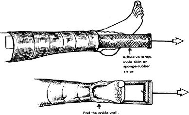
b. Check alignment of the leg to maintain a straight line of pull from the rope attached to the spreader bar to the pulley mounted on the foot of the bed. Also check the bandage wrappings and tape or moleskin strips to be sure that they are adhering properly and have not slipped downward. Report immediately if any part of the wrappings or traction apparatus appears to be out of place.
1-30. RUSSELL TRACTION
a. In this form of skin traction, a system of suspension and traction pull is used. Adhesive strips are applied as in Buck’s extension, and the knee is suspended in a sling. A rope is attached to the sling’s spreader bar. This rope passes over a pulley which is attached to an overhead bar and is then directed to a system of three pulleys at the foot of the bed: first to a pulley on the bed’s foot bar, next to a pulley attached to the foot spreader bar, and then back to a second pulley on the bed’s foot bar. There is an upward pull from the sling pulley and a forward pull from the pulleys at the foot of the bed. In Russell traction, the angle between the thigh and the bed is approximately 20° and there is always slight flexion of both the hip and the knee. The advantage of Russell traction is that some movement in bed is permissible. The patient can turn slightly toward the side in traction for back care, bedpan placement, or linen change.
b. Check the popliteal space for signs of pressure from the sling such as redness, indentations, abrasions, or pain. Check all the tape and wrappings as in Buck’s traction. Keep the patient from sliding down the bed. The foot of the bed may be elevated to help prevent this.
1-31. PELVIC TRACTION GIRDLE
a. The pelvic traction girdle is ordinarily used for treatment of low back pain and muscle spasm. It is fitted snugly and evenly over the iliac crests. The traction straps, extending on the lateral side of each thigh, are hooked to a separate rope at mid-thigh level and each rope leads to a separate but equal weight at the foot of the bed. The foot of the bed is usually elevated to provide counter traction.
b. Keep the girdle and the underlying skin clean and dry. Avoid using padding unless the patient is very thin or the iliac crests are very prominent. Protect and support the feet. Foot exercises are usually encouraged, but there must be no contact with the traction ropes. The physician’s orders may specify when the girdle may be removed for skin care or bathroom privileges.
1-32. PELVIC TRACTION SLING
a. The pelvic traction sling is used in the treatment of pelvic fracture. The patient is placed in a canvas sling or hammock that is suspended by a tension spring to an overhead frame bar. The pelvis is suspended so that it is just off the mattress.
b. Padding may be placed along the sling edges or as needed to relieve pressure on the coccyx. Keep the sling, the padding, and the skin clean and dry.
1-33. CERVICAL TRACTION HALTER
a. A canvas head halter is used for treatment of affections of the cervical spine. The halter fits snugly under the chin and around the back of the head against the occipital protuberance. A pulley rope is attached to the spreader bar that hooks to the top of the harness. The prescribed weights at the end of the pulley rope keep the patient’s neck and cervical spine in a position specified by the physician.
b. The patient’s bed may be positioned in reverse to allow easier access to the patient’s head. The head of the bed may be elevated to provide counter traction and to help prevent the patient’s head or the spreader bar from resting against the bed frame. When positioning the bed, allow for plenty of room around the head of the bed in order to prevent bumping the weights.
c. Feed the patient slowly and carefully. If turning is not permitted, remind the patient to face forward and not turn toward the spoon, fork, or straw. Allow plenty of time for him to chew and swallow. Check to be certain that the chinstrap is not pressing on his throat. More importantly, keep suction equipment on hand for immediate use to prevent the patient from aspirating when eating, drinking, or receiving mouth care. Remember, if the patient chokes or vomits, he cannot be turned to the side or raised upward.
1-34. SKELETAL TRACTION
a. Skeletal traction is used most frequently in the treatment of fractures of the femur, the tibia, the humerus, and the cervical spine. The traction is applied directly to the bone by use of a metal pin or wire inserted into or through the bone or by tongs inserted into the skull. The pin, wire, or tong is then attached to the traction apparatus.
b. A significant problem with skeletal traction is the potential for infection, which could develop in or around the insertion site. The site must be inspected daily for drainage and odor. Daily cleaning and dressing changes may be prescribed by the physician or by local standing operating procedures.
c. The insertion of pins, wires, or tongs is often done in the operating room under anesthesia. Frequently, the patient will arrive on the ward with most of the traction apparatus already in place. Assist the physician or the orthopedic technician with positioning of the patient and arrangement of the traction apparatus. Because of differences in age, weight, body type, and the nature of the fracture itself, no two fractures can be considered alike and each patient will require individualized treatment. Therefore, traction procedures are modified for the requirements of each patient. It is extremely important that nursing personnel understand the nature of the traction in use and the patient movement that is permissible while still maintaining the desired traction pull. These factors will affect the planning of basic nursing care for that patient. The following paragraphs discuss several of the most commonly used forms of skeletal traction.
1-35. CERVICAL SKELETAL TRACTION
a. Crutchfield or Vinke tongs are used for skeletal traction in the treatment of fractures of the cervical spine. The tong points are inserted in the parietal area of the skull (just in the outer layers of the bone) and the tong is then attached to the pulling device. The procedures may be done under local anesthesia in the operating room or on the ward. With skeletal skull traction, the nursing care of the patient is usually less difficult than when a halter is used–the patient’s head and face are relatively free of pressure and some turning in a “log-roll” fashion may be permissible for back care and bed making.
b. Prepare the bed as for cervical halter traction. Use an alternating pressure mattress, if one is available, when the patient is in a conventional bed. The patient in tong traction may be immobilized for a long period of time, so he may be placed on a Foster frame.
c. As with cervical halter traction, feed the patient slowly and with great care. Allow plenty of time to chew and swallow. Keep suction equipment at the bedside for emergency use.
1-36. SKELETAL TRACTION WIRE OR PIN
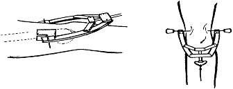
The Kirschner wire and Steinmann pin are commonly used devices in skeletal traction. The wire or pin insertion is always an aseptic procedure, and is usually done in the operating room. A local or general anesthesia is used, and all preoperative and postoperative precautions must be taken. The wire or pin is inserted through the bone, distal to the fracture site, and out through the skin on the other side. The sharp protruding ends of the pin or wire should be covered–corks are generally used for this purpose. A skeletal tractor device is fitted onto the wire or pin and traction is maintained by the weight, pulley, and rope attached to the skeletal tractor (see figure 1-15).
1-37. SKELETAL TRACTION FOR THE FEMUR
a. The combination of skeletal traction and balanced suspension is widely used for the treatment of fractures of the femoral shaft (see figure 1-16). This method of treatment provides considerable freedom of body movement while maintaining efficient traction on the injured limb. The Thomas leg splint and Pearson attachment are used to achieve this balanced suspension traction.
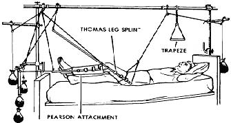
b. The Thomas splint (half ring) is applied in various ways: with the ring fitted posteriorly against the ischium or anteriorly in the groin. The thigh rests in a canvas or bandage-strip sling with the popliteal space left free. The leather ring should not be wrapped or padded. If kept smooth, dry, and polished, the leather of the ring is designed to rest against the skin and resist moisture.
c. The Pearson attachment is attached by clamps to the Thomas splint at knee level. A canvas or bandage-strip sling supports the lower leg and provides the desired degree of knee flexion. A footplate is attached to the distal end of the Pearson attachment to support the foot in a neutral position. The heel should be left free.
d. The traction is in line with the long axis of the femoral shaft and is maintained by the rope, pulley, and weights attached to the skeletal tractor, which is fitted onto the wire or pin. Counter traction and balanced suspension are provided by the ropes, pulleys, and weights attached to the Pearson attachment. When all is operational, the thigh and Thomas splint will be suspended at about a 45° angle with the bed and the lower leg and Pearson attachment will be suspended horizontal to the mattress. The patient may sit up, turn toward the traction side, and raise his hips above the bed by means of the trapeze and still maintain the line of traction.
1-38. ARM TRACTION
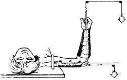
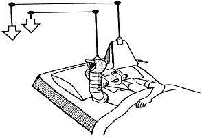
a. The type of traction used for the upper extremities will depend upon the location of the fracture, any associated injuries, and the preference of the physician. As with other body parts, the arm may be immobilized in skin traction or skeletal traction. The position of the arm in traction may be sidearm or overhead. See figures 1-17 and 1-18. On occasion, the arm may be positioned in extension. This however, will cause muscle strain and elbow joint discomfort if immobilized in this position for more than a very short period of time.
b. Nursing care considerations for the patient immobilized in arm traction are the same as for any other immobilized patient. In addition, the nursing personnel must observe the following precautions:
(1) Compare the radial pulse on the affected side with the pulse on the unaffected side. Circulatory impairment must be reported immediately.
(2) Keep the elevated hand in a position of function at all times, and observe for pressure points at the wrist. Be sure that only the fingertips extend from the sling in the overhead traction set-up.
1-39. NURSING MANAGEMENT OF THE PATIENT IN TRACTION
As stated earlier in the text, the basis of the nursing care plan will be determined by two factors: the basic position of the patient in traction and permissible movement. Normal activities of daily living are significantly altered by immobilization and confinement. Nursing management begins with assessing the patient. What are his needs? What are his limitations? Determine which activities the patient can do by himself and with which activities he requires assistance. Basic considerations are nutritional needs, hygiene, and elimination needs and the need for some sort of diversional activities. In addition to this, nursing management involves maintenance (keep traction from being compromised) and prevention (observe for complications).
a. When assisting with a.m. and p.m. care, encourage the patient to do as much for himself as is possible within the constraints of his immobilization. Assist with or perform those tasks that the patient cannot perform.
b. Assess the patient and the traction set-up to determine the best method for changing the bed linen. There are several acceptable methods for making an occupied bed and, depending upon the type of traction in use, you will want to use the method that is easiest. For some patients, a head-to-toe technique may work better than side-to-side. Always be sure that the linen is smooth and dry. Utilize draw sheets when appropriate. Reposition supporting pillows and change the pillow cases as often as needed to prevent the patient from being supported by soiled, damp, wrinkled, or flattened pillows.
c. When assisting with the bedpan or urinal, provide adequate time and privacy for the patient. Many patients do not adjust easily to the awkwardness of using a bedpan or urinal. The presence of roommates, visitors, or hospital personnel just outside the privacy curtain is enough to make anyone uncomfortable. Always place toilet tissue, moist towelettes, and call bell within easy reach. Check daily to see whether the patient has had a bowel movement. Treating constipation will prevent the more serious problem of fecal impaction. Physicians will routinely prescribe a stool softener for immobilized patients in order to prevent constipation.
d. Encourage the patient to eat all of the prescribed diet. If permitted by the physician, suggest that family and friends bring fruit or a “healthy” favorite food from home. A recovering patient’s diet should be high in calcium, protein, iron, and vitamins. Plenty of fluids and foods high in roughage will help prevent bowel and bladder complications.
e. Assist the patient to take several deep breaths each hour. Coughing and deep breathing will help prevent respiratory complications. Encourage the patient to actively exercise the unaffected extremities.
f. Eliminate any factors that reduce the traction pull or alter its direction. Ropes and pulleys should be in straight alignment and the ropes should be unobstructed. Traction is NOT accomplished if the knot in the rope is touching the pulley or the foot of the bed. The weights must be suspended and not in contact with the bed or resting on the floor. The patient’s body should always be in alignment with the force of traction. Check the patient’s position each time you enter the room and help the patient slide up in bed if necessary. Encourage the patient to use the overhead trapeze instead of elbows to move in bed.
g. Check the extremities for color (pallor, cyanosis), numbness, edema, signs of infection, and pain. Look for areas of skin breakdown or pressure sores on all skin surfaces.
h. Orthopedic patients confined in traction will need some sort of diversional activity to relieve boredom and prevent depression. If your treatment facility has no occupational therapy department, encourage family and friends to visit frequently and bring books or games for the patient. Television and radio may also help to pass the time. The nursing personnel should make opportunities to stop and chat with the patient, both to distract the patient from boredom and to assess the patient’s mental status. It is often easy to see a state of depression beginning and it will be easier to dispel in its early stages.
