2-1. INTRODUCTION
a. The respiratory tract is the most common portal of entry and exit of microscopic disease agents. Many of these microorganisms leave the body of the infected person by means of droplets and by nose and throat secretions. Droplets are exhaled in coughing, sneezing, talking, or simply breathing. These droplets do not always fall to the ground immediately, but may remain suspended in the air for many hours and can be inhaled by a well person, who may then become infected. The infection may also be spread to a well individual who improperly handles secretions of the nose and throat of an infected person. Many respiratory diseases are infectious in nature and are easily spread.
b. Medical intervention and skilled nursing care are employed in treating respiratory infections. Skilled nursing care includes knowledge of the duration and stages of the disease, isolation procedures, infection control policies, comfort measures for the patient, therapeutic measures, and observation of signs, symptoms, and potential complications.
2-2. THE RESPIRATORY SYSTEM
a. The cells of the body require a constant supply of oxygen to carry on the chemical processes necessary to life. As a result of these processes, carbon dioxide, a waste product, is formed, and must be removed from the body. Oxygen and carbon dioxide are continually being exchanged, both between the body and the atmosphere and within the body. This process is known as respiration, and the body system that performs this exchange of gases is the respiratory system.
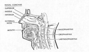
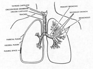
b. The respiratory system is a continuous series of passages that begins with the nose and ends in the alveoli of the lungs. The upper respiratory system includes the nose, pharynx, larynx, and the trachea. The lower respiratory system includes the right and left bronchi, their subdivisions, and the lungs. Review Figure 2-1 as you read the next paragraph.


2-3. STRUCTURE AND FUNCTION
a. Nose. Inhaled air is warmed, moistened, and filtered in the nasal cavities. Filtering is done by the cilia (tiny hair-like projections) of the mucous membrane lining the nasal passages.
b. Pharynx. The pharynx, or throat, connects the nose and mouth with the lower air passages and the esophagus. It is divided into three sections, the nasopharynx, the oropharynx, and the laryngopharynx. Both air and food pass through the pharynx. Air passes from the nose and mouth to the larynx, while food passes from the mouth to the esophagus. The walls of the pharynx contain masses of lymphoid tissue called the adenoids and tonsils.
c. Larynx. The larynx, or voice box, connects the pharynx with the trachea. Two membranous bands in the wall of the larynx are called vocal cords. Vibrations of the vocal cords produce sound.
d. Trachea. The trachea, or windpipe, is a tube that carries air from the larynx to the bronchi. It is held open by cartilaginous rings and is lined with cilia and mucous glands to keep dust and dirt out of the lungs.
e. Bronchi. The trachea divides to form the two main bronchi. One bronchus enters each lung and divides into many smaller air passages called bronchioles. The bronchioles terminate in the final air spaces, called the alveoli.
f. Lungs.
(1) The lungs are the organs of respiration. They are elastic structures contained within the thoracic cavity. The upper, pointed border of each lung, called the apex, extends above the clavicle. The lower border, or base, rests upon the diaphragm. Each lung is divided into sections called lobes. The right lung has three lobes, termed upper, middle, and lower. The left lung has only two lobes, referred to as upper and lower. Inside each lung, millions of tiny air sacs, called alveoli, are interlaced in a network of capillaries. Certain cells in the alveolar walls secrete a lipid-rich material called surfactant. Surfactant helps to maintain the elastic quality of the alveolar membrane and assists with the transfer of gases.
(2) Oxygen-poor blood is pumped from the heart’s right ventricle, through the right and left pulmonary arteries, and into the lungs. Paralleling the branching of the respiratory tree, the arteries divide and subdivide within the lungs. Arteries divide into arterioles, and arterioles divide into the capillaries that surround the alveoli.
(3) Each lung is enclosed by a membranous sac formed of two layers of serous membrane called pleura. One layer covers the lungs and is called the visceral pleura. The other lines the chest cavity and is called the parietal pleura. The space between the two layers, the pleural cavity, contains a small amount of fluid that lubricates the surfaces. Between the two lungs is the mediastinum, the central thoracic cavity containing the heart, the great vessels, the esophagus, and the lower trachea.
2-4. PHYSIOLOGY OF RESPIRATION
a. The walls of the alveoli are composed of a thin, permeable membrane. It is here that oxygen passes from the alveoli into the tiny capillaries that surround each alveoli. Carbon dioxide in the blood passes from the capillaries into the alveoli to be exhaled. This exchange of oxygen and carbon dioxide in the lungs is called external respiration.
b. The oxygen that enters the capillaries is carried by the red blood cells in a chemical combination with hemoglobin. This blood, now oxygenated, returns to the heart to be pumped out to the body through the arteries. The oxygen passes part of the blood into the cells of the body, while carbon dioxide waste from the cells is passed into the blood that returns to the heart. This exchange of gases between the capillary blood and the cells of the body is called internal respiration.
2-5. MECHANICS OF RESPIRATION
a. The act of breathing, the cycle of inspiration and expiration, is repeated about 16-20 times per minute in a resting adult. Breathing is regulated by the respiratory center in the medulla of the brain. The level of carbon dioxide (CO) in the circulating blood is one of the major influences upon the respiratory reflex. The respiratory center is sensitive to changes in blood composition, temperature, and pressure, and will adjust the rate and depth of breathing to accommodate the body’s needs.
b. The physical conditions that control the flow of air into and out of the lungs are referred to as the mechanics of ventilation. Air flows from an area of higher pressure to an area of lower pressure. During inspiration, contraction of the diaphragm and intercostal muscles increases the size of the thoracic cavity. This causes the pressure within the thoracic cavity to become less than that of the atmosphere, and air is drawn through the air passages into the alveoli. During normal expiration, relaxation of the same muscles will cause the thoracic cavity to decrease in size, thereby increasing the pressure within the thoracic cavity to that which is greater than atmospheric pressure. Air will then flow out of the lungs into the atmosphere.
