This is the Archived Desktop Edition.
You should be transferred to the Newest Edition for Desktop and Mobile within 2 seconds.
![]()
Multimedia Edition
|
Lesson 2: Embryology and Fetal Development
2-11. COMPONENTS OF FETAL CIRCULATION
As the placenta acts as the intermediary organ of transfer between the mother and fetus, fetal circulation differs from that required for extrauterine existence. The fetus receives oxygen through the placenta because the lungs do not function as organs of respiration in the uterus. To meet this situation, the fetal circulation contains certain special vessels that shunt the blood around the lungs, with only a small amount circulating through them for nutrition. See figure 2-9. The following functions occurs:
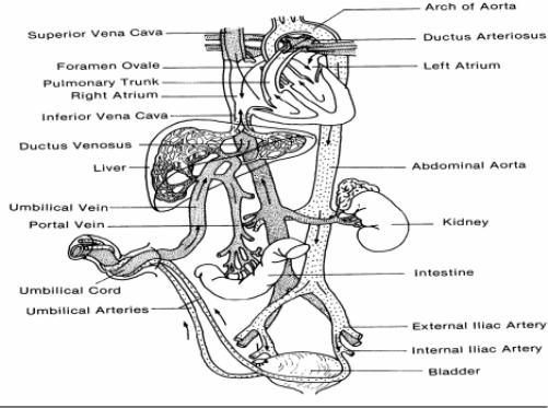
Figure 2-9. Fetal
circulation before birth.
a. The umbilical vein transports blood rich in oxygen and nutrients from the placenta to the fetal body. This vein travels along the anterior abdominal wall of the fetus to the liver, and at the porta hepatis, the umbilical vein divides into two branches.
b. About 1/2 of the blood passes into the liver and the rest enters a shunting vessel called the ductus venosus that bypasses the liver. The ductus venosus travels a short distance and joins the inferior vena cava.
c. There, the oxygenated blood from the placenta is mixed with deoxygenated blood from the lower parts of the fetal body. This blood continues through the vena cava to the right atrium.
d. As the blood relatively high in oxygen enters the right atrium of the fetal heart, a large proportion of it is shunted directly into the left atrium through an opening in the atrial septum called the foramen ovale.
e. The more highly oxygenated blood that enters the left atrium through the foramen ovale is mixed with a small amount of deoxygenated blood returning from the pulmonary veins. This mixture moves into the left ventricle and is pumped into the aorta.
f. Some of this blood reaches the myocardium by means of the coronary arteries and some reaches the tissues of the brain through the carotid arteries.
g. The rest of the blood entering the right atrium, as well as the large proportion of the deoxygenated blood entering from the superior vena cava, passes into the right ventricle and out through the pulmonary artery.
h. Enough blood reaches the lung tissues to sustain them.
i. Most of the blood in the pulmonary artery bypasses the lungs by entering the ductus arteriosus, which connects the pulmonary artery to the descending portion of the aortic arch.
j. Some of the blood carried by the descending aorta leads to the various parts in the lower regions of the body.
k. The rest of the blood passes into the umbilical arteries which branch from the internal iliac arteries and lead to the placenta.
2-12. CHANGES CONTINUE IN CIRCULATION AFTER BIRTH
See figure 2-10.
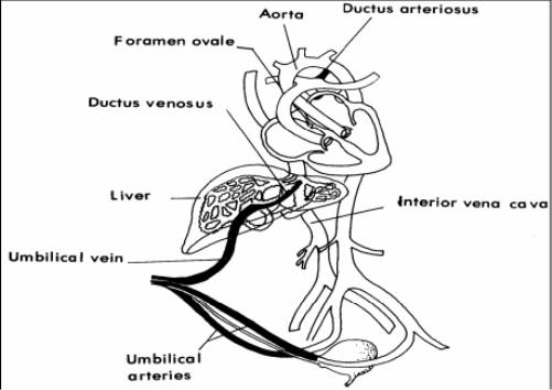
Figure 2-10. Fetal
circulation after birth.
a. The umbilical vein is obliterated and becomes the round ligament of the liver.
b. The umbilical arteries are obliterated and ultimately become ligaments.
c. The ductus venosus is obliterated and becomes a ligament. Anatomic closure is completed at the end of 2 months. The ductus venosus is superficially embedded in the wall of the liver.
d. The ductus arteriosus is obliterated and becomes a ligament. Functional closure takes 3-4 days; anatomic closure is completed by 3 weeks. The constriction seems to be stimulated by a substance called Bradykinin, which is released from the lungs during their initial expansions.
e. The foramen ovale closes after the umbilical cord is tied and cut. A large amount of blood is returned to the heart and the lungs. With the lungs now functioning, there is equal pressure on both atria as the vessels constrict. Failure of the foramen ovale to close spontaneously results in an atrial septal defect, which may or may not require surgery later.
|
|
|
|
The Brookside Associates Medical Education Division is dedicated to the development and dissemination of medical information that may be useful to medical professionals and those in training to become medical professionals. This website is privately-held and not connected to any governmental agency. The views expressed here are those of the authors, and unless otherwise noted, do not necessarily reflect the views of the Brookside Associates, Ltd., any governmental or private organizations. All writings, discussions, and publications on this website are unclassified.
© 2007 Medical Education Division, Brookside Associates, Ltd. All rights reserved
![]()