This is the Archived Desktop Edition.
You should be transferred to the Newest Edition for Desktop and Mobile within 2 seconds.
![]()
Multimedia Edition
|
Lesson 2: Embryology and Fetal Development
The placenta is a fleshy disk like organ. The fully developed placenta (afterbirth) is reddish in color. It is formed from the outer layers of the blastocyst. It is completely formed by the third month of pregnancy. The umbilical cord (lifeline) connects the fetus to the placenta and is normally 20 inches in length and 3/4 inch in diameter. It contains one umbilical vein and two umbilical arteries.
2-6. FUNCTIONS OF THE PLACENTA
Being knowledgeable of the placenta functions gives insight into prenatal life and is helpful in providing nursing care to the unborn and the newborn. The placenta functions as a transport mechanism between the embryo and the mother (see figure 2-6). The placenta has many tasks: it transports oxygen, nutrients, and antibodies to the fetus by means of the umbilical vein; removes carbon dioxide and metabolic wastes from the fetus by the two umbilical arteries; serves as a protective barrier against harmful effects of certain drugs and microorganisms; acts as a partial barrier between the mother and fetus to prevent fetal and maternal blood from mixing; and produces hormones essential for maintaining the pregnancy. (The hormones are estrogen, progesterone, and human chorionic gonadotropin (HCG)).
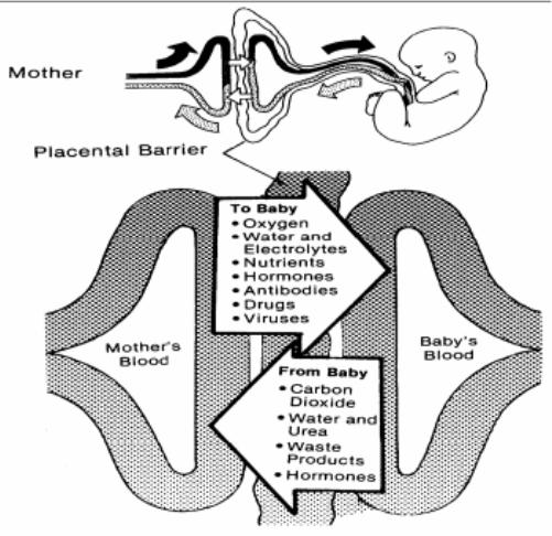
Figure 2-6. The placental circulation.
Two closely applied but separate membranes line the uterine cavity and surround the developing embryo-fetus. Both membranes, the amnion (inner membrane) and the chorion (outer membrane), arise from the zygote. As the chorion develops, it blends with the fetal portion of the placenta; the amnion blends with the fetal umbilical cord. These deceptively strong, translucent membranes contain not only the fetus but also the amniotic fluid, and they are continuous with the margins of the placenta. See figure 2-7.
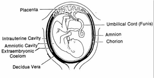
a. Amnion. This is the smooth, slippery, glistening innermost membrane that lines the amniotic space. It is filled with fluid and is often called the "bag of water." The fetus floats and moves in the amniotic cavity. At full term, this cavity normally contains 500 cc to 1000 cc of fluid (water). This fluid provides many functions for the fetus. The amnion usually ruptures just before birth. The amnion functions to:
(1) Protect the fetus from direct trauma by distributing and equalizing any impact the mother may receive.
(2) Separate the fetus from the fetal membranes.
(3) Allow freedom of fetal movement and permits musculoskeletal development.
(4) Facilitate symmetric growth and development of the fetus.
(5) Protect the fetus from the loss of heat and maintains a relative, constant fetal body temperature.
(6) Serve as a source of oral fluid for the fetus.
(7) Act as an excretion and collection system.
b. Chorion. This is the outer membrane. It forms a large portion of the connective tissue thickness of the placenta on its fetal side. It is the structure in and through which the major branching umbilical vessels travel on the surface of the placenta.
|
|
|
|
The Brookside Associates Medical Education Division is dedicated to the development and dissemination of medical information that may be useful to medical professionals and those in training to become medical professionals. This website is privately-held and not connected to any governmental agency. The views expressed here are those of the authors, and unless otherwise noted, do not necessarily reflect the views of the Brookside Associates, Ltd., any governmental or private organizations. All writings, discussions, and publications on this website are unclassified.
© 2007 Medical Education Division, Brookside Associates, Ltd. All rights reserved
![]()