|
Evaluate the pelvis systematically.
Position the patient at the very edge of the exam table, with her
feet in stirrups, knees bent and relaxed out to the side. If she is not
down far enough, the exam will be more difficult for you and more
uncomfortable for her.
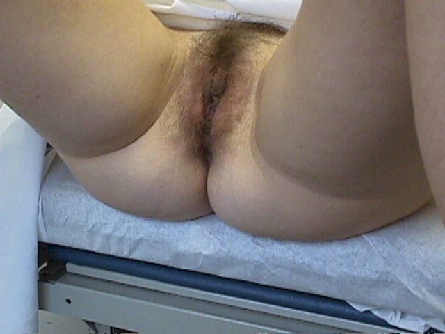
Position the patient at the edge
of the exam table.
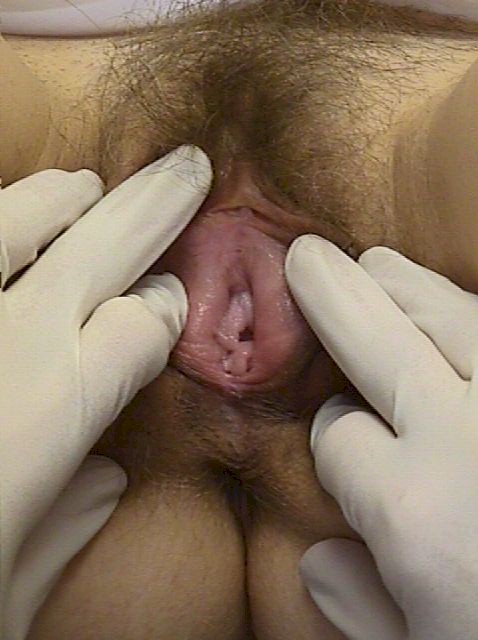
Inspect the vulva.
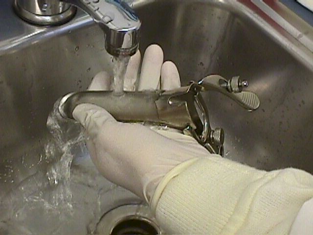
Warm and lubricate the speculum .
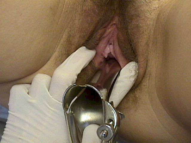
Separating the labia with one hand,
insert a warmed, water-lubricated
speculum with the other hand.
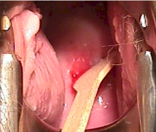
Obtain a Pap smear or cervical
cultures,
if indicated.
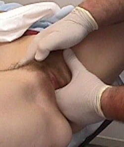
Feel for masses or tenderness. |
Pad the stirrups to avoid the stirrups digging into her feet. Kitchen
pot-holders work well for this, but almost any soft material can be
used.
Use a bright light to visually inspect the vulva, vagina and cervix.
Most examiners find it easiest to look just over the light to get the
best view. Separate the labia with your gloved fingers to look for any
surface lesions, redness, or swellings. Look within the pubic hair for
the tiny movement of pubic lice or nits. Look on the labia for the
cauliflower-like bumps that are known as venereal warts. Using
magnification (magnifying lenses or colposcope) is very useful when the
patient has vulvar complaints and the diagnosis is not obvious.
Look between the folds of skin for ulcerative lesions that can
indicate an active herpes infection. Gently retract the clitoral hood
back, exposing the clitoris while looking for peri-clitoral lesions.
Look for the hymen or remnants of the hymen and identify any redness
just exterior to the hymen that can indicate vulvar vestibulitis.
The periurethral glands (Skene's glands) have tiny ducts that open
onto the surface. Look for them next to the urethra. While looking at
the urethra, note any discharge coming from the urethral opening that
might suggest gonorrhea or chlamydia.
Palpate the upper labia majora for masses related to hernias
extending through the Canal of Nuck. Palpate the middle and lower
portion of the labia majora for masses suggesting a Bartholin Duct Cyst.
After warming a vaginal speculum with warm water, separate the labia
with one hand while gently inserting the speculum with the other hand.
It is frequently more comfortable for the patient if you insert the
speculum rotated about 45 degrees (so the blades are not horizontal but
are oblique). Once past the introitus, rotate the speculum back to it's
normal position.
The labia, particularly the labia minora, are very sensitive to
stretching or pinching, so try not to catch the labia minora in the
speculum while inserting it.
Some gynecologists ask their patients to "bear down" while they are
inserting the speculum and feel that this assists with insertion. Others
find this instruction to be be confusing and don't use it.
Obtain specimens for a Pap smear and any
cultures that may be indicated.
Then feel the pelvis by application of a "bimanual exam." For a normal
examination:
-
External genitalia are of normal appearance. There is no enlargement of the Bartholin or
Skene glands.
-
Urethra and bladder are non-tender.
-
Vagina is clean, without lesions or discharge
-
Cervix is smooth, without lesions. Motion of the cervix causes no pain.
-
Uterus is normal size, shape, and contour. It is non-tender.
-
The adnexa (tubes and ovaries) are neither tender nor enlarged.
Pelvic Anatomy Video
During the bimanual exam, you may use one finger or two fingers
inside the vagina. Two fingers allows for deeper penetration and more
control of the pelvic structures, but one finger is more comfortable for
the patient. You should individualize your exam for the specific
patient.
Turning your hand palm up, compress the urethra against the underside
of the pubic bone. Normally, this doesn't hurt. If it causes discomfort
for the patient, it is likely that at least some degree of urethritis is
present.
Then insert your fingers deeper into the pelvis. Keeping your palm
up, curl your vaginal finger(s) up, compressing the bladder against the
back of the pubic bone. Normally, this pressure creates the sensation
that the patient needs to urinate, but is not painful. If it is painful,
this is good clinical evidence of cystitis (urinary tract infection), or
(less likely) endometriosis.
In some patients, particularly those with difficult to feel pelvic
masses, a combined rectovaginal exam is useful. Change gloves, lubricate
the rectum, and then gently insert your index finger into the vagina and
your middle finger into the rectum. The rectovaginal exam is helpful in
feeling the uterosacral ligaments, a common site of endometriosis
involvement.
On completion of the rectal exam, stool can be checked for the
presence of occult blood.
If the hymen is intact, it may still be possible to perform a
comfortable and complete exam, but if the exam is causing too much pain,
stop the exam and consider these alternatives:
-
Rectal exam with your index finger can often provide all the
information you need at that time.
-
Exam under anesthesia will provide full access without causing
pain to the patient.
-
Ultrasound scan, abdominally and trans-perineal, can sometimes
provide you with the information you need.
|


