Other Services
Cultures · Rectal
· Urine · Wet Mount
· Mammogram · Breast Self Exam
· Counseling · Plan
· Charting
Cultures
Cultures can sometimes be helpful in determining the cause for vaginal or vulvar
symptoms such as pain, burning or itching. The cultures can be in addition to
a wet mount, or supplementary to a wet mount.
Bacterial cultures for
Strep, E. coli and other pathogens may then indicate a course
of treatment that would not necessarily be obvious from either the gross
appearance of the vagina or the wet mount.
Some physicians routinely culture for gonorrhea and/or chlamydia on all of their
patients at each routine visit. Others culture for these STDs only among
high risk patients or those with unexplained pelvic pain. The wisest
course for you depends on the frequency with
which these STDs are found in your population.
Rectal
While some physicians routinely perform a rectal exam on all patients, others perform a
rectal only on selected individuals in certain clinical circumstances, such as after age
50.
Routine screening with sigmoidoscopy every 5 years after age 50 is recommended by many
physicians.
After the rectal exam, the small particles of stool left on the examining glove can be
evaluated for the presence of occult blood. This is most useful after the age of 50.
Urine
Some physicians routinely check the urine at each routine visit.
Others check the urine only for a specific indications. A clean urine specimen can be
evaluated for the presence of:
Wet Mount
Vaginal discharge can be evaluated using a "wet mount."
Mix a small amount of discharge with 10% potassium hydroxide (KOH), place
it on a glass slide and cover it with a coverslip. The KOH dissolves cell membranes, making it
easier to see yeast organisms under the microscope.
Mix another small amount of discharge with a drop of normal saline, place
it on a
glass slide with a coverslip, and examine it under the microscope. With saline, active trichomonad organisms
can be seen moving and "clue cells," indicating bacterial vaginosis can be seen.
Wet mount Video.
Read more about performing a wet mount.
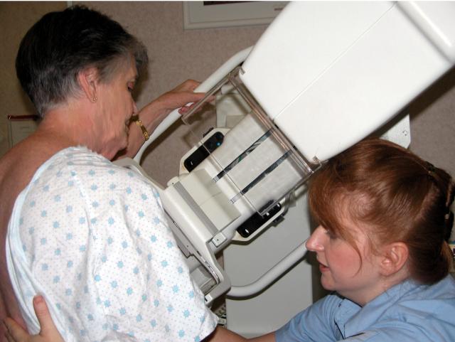
Mammography
 Mammography is a useful method of evaluating the breasts for the possible presence of
early malignancy. Mammography is a useful method of evaluating the breasts for the possible presence of
early malignancy.
While not 100% accurate, mammography is probably around 80% accurate, particularly in detecting
the very small, early malignancies not appreciated by physical examination.
Recommendations for frequency of mammograms vary, but the following general guidelines can
be followed:
-
Women with a disquieting symptom (eg bloody nipple discharge) or physical finding may
benefit from an indicated mammogram.
-
Women with no significant high risk factors will probably benefit from routine mammogram
screening every other year, from age 40 to 50, and annually after age 50.
-
Women with a strong family history of breast cancer or other significant high risk
factor may benefit from more frequent mammogram screening, and starting at a younger age.
Read the breast cancer screening chapter from the Guide to Clinical Preventive
Services, Second Edition, Report of the U.S. Preventive Services Task Force.
Breast Self-examination
An important part of patient education is to see that she feels confident in her skills
at self-breast examination. If not, you can teach her the proper techniques. I sometimes
inquire:
"Are you examining your breasts regularly?"
Many offices use a video to demonstrate breast examination
techniques. Some use a manikin with several breast lumps for the
patient to identify. Some have a patient information sheet the woman
can take home to study on her own.
Counseling
Counseling may be brief or lengthy.
It may be focused on the problems presented during the examination, or may be global,
such as diet, exercise, or other healthy life-styles.
Patients often feel this is the most important part of the visit. Take your time and
sit down while talking to the patient. You need not be a master of "bed side
manner" for the patient to appreciate this time. Just be honest, direct, and
pleasant.
Plan
Before leaving, the patient should understand any future plans.
Laboratory requisitions or consultation requests can be given. Patient hand-outs can be
provided. Plans might include:
It is routine to indicate when the patient should return to the office (RTO) or return
to the clinic (RTC).
"RTO in _______ months."
Charting
Many health care providers find it useful to utilize a standard form for recording
information about this exam. An example is shown here:
Office Exam
|


