a. Testes. The testes are the primary organs of reproduction in the male.
The male testes correspond to the female ovaries.
(1) Description/information. The testes are located in the scrotum.
They are oval structures enclosed in a fibrous capsule. The testes are covered by a dense layer of white fibrous tissue called the tunica albuginea. This tissue layer extends inward and divides each testis into a series of internal compartments called lobules. Each of the 200 to 300 lobules contains one to three tightly coiled tubules called the seminiferous tubules.
(2) Functions. The seminiferous tubules produce sperm by a process called spermatogenesis.
As well as producing sperm, the testes produce the male hormone testosterone. Interstitial cells within the testes produce this hormone, which is essential for the development of the male secondary sex characteristics. If testosterone is not produced in a male body, growth of hair on the face and body, deepening of the voice, and an increase in skeletal mass do not occur. Also, sperm will not develop without testosterone.
(3) Sperm. The seminiferous tubules produce sperm by a process called spermatogenesis. Sperm can be defined as the reproductive cells of the male. Each seminiferous tubule is packed with sperm in various stages of development. Beginning at about puberty, a male produces about 300 million sperm cells each day. As a male grows older, the production of sperm decreases. Males continue to produce sperm throughout life.
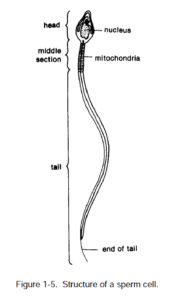
(a) Description/information. Compared to a female ovum, a sperm cell is very small, but it is well shaped to reach out and penetrate a female ovum. A sperm cell has a head, a middle section, and a tail. The head is flat and oval shaped (ideal for penetration and attachment) and contains the nucleus of the cell. The middle section is made up of substances that make useable energy to propel the tail. And the long tail acts like a whip to move the sperm. When the head penetrates the ovum, the tail separates from the rest of the sperm.
(b) Chromosomes in a sperm cell. The nucleus in the head of a sperm cell contains chromosomes. A mature sperm has 23 chromosomes. An immature sperm cell has 46 chromosomes, one an X (female) chromosome and the other a Y (male) chromosome. A reduction division takes place to form a mature cell which has 23 chromosomes. At that time, an X chromosome (female) goes to one sperm cell, and a Y (male) chromosome goes to the other sperm cell. If an ovum is joined by a sperm with an X chromosome, the combination will form a female. If a sperm with a Y chromosome joins an ovum, a male is formed.
b. Epididymis. At the upper and posterior part of each testis is the epididymis–an elongated, triangular tube which is 16 to 20 feet in length.
Each comma-shaped tube is positioned along the posterior side of a testis and is mostly made up of a tightly coiled tube called the ductus epididymis. Sperm mature in the epididymis tubes. These tubes link the testes proper with the ductus deferens. Sperm are stored in the epididymis tubes until they are ejaculated and enter the vas deferens.
c. Ductus (Vas) Deferens. At its tail, the epididymis becomes less coiled, its diameter increases, and the tubes become known as the ductus deferens or the vas deferens.
Ductus deferens are muscular tubes which are about 48 centimeters (18 inches) long. Two ductus deferens, one from each epididymis tube, lead up through the inguinal canal into the pelvic cavity, cross to the posterior surface of the urinary bladder, and unite with the ducts of the seminal vesicles to form the ejaculatory ducts. Each ductus deferens stores sperm for a period of up to several months and propels sperm toward the urethra during ejaculation.
d. Seminal Vesicles. The seminal vesicles are two glandular pouches located behind and below the urinary bladder.
These tubular structures secrete a fluid which activates the spermatozoa in the semen. The secretions contain sugar fructose and prostaglandins. Fructose energizes the sperm, and prostaglandins assist ejaculation and stimulate uterine contractions. Thus, both fructose and prostaglandins help sperm move to the uterine tubes where fertilization occurs. Additionally, this fluid is slightly alkaline, which helps protect sperm against the acid secretion of the vagina. Secretion of the seminal vesicles makes up 60 percent of the ejaculate (fluid ejaculated).
e. Ejaculatory Duct. Each ductus deferens and its corresponding seminal vesicle come together to form a short tube called the ejaculatory duct.
The ejaculatory duct opens into the urethra within the prostate gland. The ejaculatory duct carries both sperm and seminal vesicle fluid.
f. Prostate Gland. This gland is a single, doughnut-shaped gland which is about the size of a chestnut.
The gland lies directly below the urinary bladder and surrounds the prostatic part of the urethra. The prostate gland secretes a highly alkaline fluid which protects sperm acidity in the urethra and vagina. Secretion from the prostate gland is added to the sperm and seminal vesicle fluid. From 13 to 33 percent of the volume of semen seminal vesicle fluid is prostate gland secretion. Prostate gland secretion also contributes to sperm motility.
g. Bulbourethral (Cowper’s) Glands.
(1) Description/information. These are two small glands, about the size of peas, located just below the prostate on either side of the urethra. These glands secrete a mucous-like lubricating fluid into the membranous urethra. The glands also secrete a substance that neutralizes urine. Ducts of these glands open into the spongy urethra.
(2) Semen. Semen (seminal fluid) is the fluid discharged at ejaculation by a male. This fluid is made up of sperm in the secretions of the seminal vesicles, the prostate gland, and the bulbourethral glands.
h. Urethra. The urethra is the final duct of the reproductive system.
This duct acts as a passageway for sperm or urine. The urethra is about 20 cm (8 inches) long.
The ejaculatory ducts pass sperm into the urethra which passes through the prostate gland and through the penis to be ejaculated.

