Basic Human Anatomy
Lesson 6: The Human Digestive System
Welcome back. Today, we embark on Lesson 6 where we’ll study the human digestive system.
After completing this lesson, you should be able to define the human digestive system, and name six major organs.
I want you to be able to name and describe six structures of the oral complex.
I would expect you to be able to describe the pharynx, the esophagus, the stomach, the small intestines, the liver and gallbladder, the pancreas, and the large intestines.
INTRODUCTION
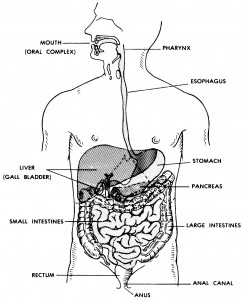
The human digestive system is a group of organs designed to take in foods, initially process foods, digest the foods, and eliminate unused materials of food items. It is a hollow tubular system from one end of the body to the other end.

Figure 6-1. The human digestive system.
Major Organs. There are eight major organs involved in the human digestive system and I’ll discuss each of them later in this lesson. The eight are:
Mouth or oral complex.
Pharynx.
Esophagus.
Stomach.
Small intestines and associated glands.
Large intestines.
Rectum.
Anal canal and anus.
Digestive Enzymes. A catalyst is a substance that accelerates (speeds up) a chemical reaction without being permanently changed or consumed itself. A digestive enzyme serves as a catalyst, aiding in digestion. Digestion is a chemical process by which food is converted into simpler substances that can be absorbed or assimilated by the body. Enzymes are manufactured in the salivary glands of the mouth, in the lining of the stomach, in the pancreas, and in the walls of the small intestine.
FOODS AND FOODSTUFFS
Examples of food items are a piece of bread, a pork chop, and a tomato. Food items contain varying proportions of foodstuffs. Foodstuffs are the classes of chemical compounds which make up food items. The three major types of foodstuffs are carbohydrates, lipids (fats and oils), and proteins. Food items also contain water, minerals, and vitamins.
THE SUPRAGASTRIC STRUCTURES
Supragastric means those structures above the level of the stomach.
ORAL COMPLEX
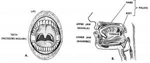
The oral complex consists of the structures commonly known together as the mouth. It takes in and initially processes food items. See figure 6-2.

Figure 6-2. Anatomy of the oral complex.
Teeth.
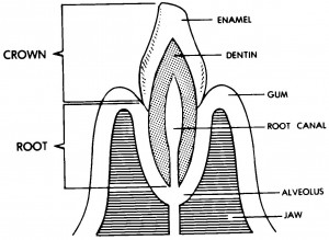
A tooth (figure 6-3) has two main parts–the crown and the root. A root canal passes up through the central part of the tooth. The root is suspended within a socket (called the alveolus) of one of the jaws of the mouth. The crown extends up above the surface of the jaw. The root and inner part of the crown are made of a substance called dentin. The outer portion of the crown is covered with a substance known as enamel. Enamel is the hardest substance of the human body. The nerves and blood vessels of the tooth pass up into the root canal from the jaw substance.

Figure 6-3. Section of a tooth and jaw.
There are two kinds of teeth– anterior and posterior. The anterior teeth are also known as incisors and canine teeth. The anterior teeth serve as choppers. They chop off mouth-size bites of food items. The posterior teeth are called molars. They are grinders. They increase the surface area of food materials by breaking them into smaller and smaller particles.
Humans have two sets of teeth–deciduous and permanent. Initially, the deciduous set includes 20 baby teeth.
DECIDUOUS = to be shed
These are eventually replaced by a permanent set of 32.
Jaws. There are two jaws–the upper and the lower. The upper is called the maxilla. The lower is called the mandible.
In each jaw, there are sockets for the teeth. These sockets are known as alveoli. The bony parts of the jaws holding the teeth are known as alveolar ridges.
The upper jaw is fixed to the base of the cranium. The lower jaw is movable. There is a special articulation (T-MJ–temporo-mandibular joint) with muscles to bring the upper and lower teeth together to perform their functions.
Palate. The palate serves as the roof of the mouth and the floor of the nasal chamber above. Since the anterior two-thirds is bony, it is called the hard palate. The posterior one-third is musculo-membranous and is called the soft palate. The soft palate serves as a trap door to close off the upper respiratory passageway during swallowing.
Lips and Cheeks. The oral cavity is closed by a fleshy structure around the opening. Forming the opening are the lips. On the sides are the cheeks.
Tongue. The tongue is a muscular organ. The tongue is capable of internal movement to shape its body. It is moved as a whole by muscles outside the tongue. Interaction between the tongue and cheeks keeps the food between the molar teeth during the chewing process. When the food is properly processed, the tongue also initiates the swallowing process.
Salivary Glands. Digestion is a chemical process which takes place at the wet surfaces of food materials. The chewing process has greatly increased the surface area available. The surfaces are wetted by saliva produced by glands in the oral cavity. Of these glands, three pairs are known as the salivary glands proper.
Taste Buds. Associated with the tongue and the back of the mouth are special clumps of cells known as taste buds. These taste buds literally taste the food. That is, they check its quality and acceptability.
PHARYNX
The pharynx (pronounced “FAIR -inks”) is a continuation of the rear of the mouth region, just anterior to the vertebral column (spine). It is a common passageway for both the respiratory and digestive systems.
ESOPHAGUS
The esophagus is a muscular, tubular structure extending from the pharynx, down through the neck and the thorax (chest), and to the stomach. During swallowing, the esophagus serves as a passageway for the food from the pharynx to the stomach. Basic Human
THE STOMACH
STORAGE FUNCTION
The stomach is a sac-like enlargement of the digestive tract specialized for the storage of food. Since food is stored, a person does not have to eat continuously all day. One is freed to do other things. The presence of valves at each end prevents the stored food from leaving the stomach before it is ready. The pyloric valve prevents the food from going further. The inner lining of the stomach is in folds to allow expansion.
DIGESTIVE FUNCTION
While the food is in the stomach, the digestive processes are initiated by juices from the wall of the stomach. The musculature of the walls thoroughly mixes the food and juices while the food is being held in the stomach. In fact, the stomach has an extra layer of muscle fibers for this purpose.
When the pyloric valve of the stomach opens, a portion of the stomach contents moves into the small intestine.
THE SMALL INTESTINES AND ASSOCIATED GLANDS
Digestion is a chemical process. This process is facilitated by special chemicals called digestive enzymes. The end products of digestion are absorbed through the wall of the gut into the blood Basic Human Anatomy Lesson 6: The Human Digestive System Page 7
vessels. These end products are then distributed to body parts that need them for growth, repair, or energy.
There are associated glands–the liver and the pancreas–which produce additional enzymes to further the process.
Most digestion and absorption takes place in the small intestines.
ANATOMY OF THE SMALL INTESTINES
The small intestines are classically divided into three areas– the duodenum, the jejunum, and the ileum. The duodenum is C-shaped, about 10 inches long in the adult. The duodenum is looped around the pancreas.
DUODENUM = 12 fingers (length equal to width of 12 fingers)
The jejunum is approximately eight feet long and connects the duodenum and ileum.
The ileum is about 12 feet long. The jejunum and ileum are attached to the rear wall of the abdomen with a membrane called a mesentery. This membrane allows mobility and serves as a passageway for nerves and vessels (NAVL) to the small intestines.
JEJUNUM = empty
ILEUM = lying next to the ilium (bone of the pelvic girdle; PELVIS = basin)
The small intestine is tubular. It has muscular walls which produce a wave-like motion called peristalsis moving the contents along. The small intestine is just the right length to allow the processes of digestion and absorption to take place completely.
The inner surface of the small intestine is NOT smooth like the inside of new plumbing pipes. Rather, the inner surface has folds (plicae). On the surface of these plicae are finger-like projections called villi (villus, singular). This folding and the presence of villi increase the surface area available for absorption.
LIVER AND GALLBLADDER
Liver Anatomy. The liver is a large and complex organ. Most of its mass is on the right side of the body and within the lower portion of the rib cage. Its upper surface is in contact with the diaphragm.
Liver Functions. The liver is a complex chemical factory with many functions. These include aspects of carbohydrate, protein, lipid, and vitamin metabolism and processes related to blood clotting and red blood cell destruction. Its digestive function is to produce a fluid called bile or gall.
Gallbladder. Until needed, the bile is stored and concentrated in the gallbladder, a sac on the inferior surface of the liver. Fluid from the gallbladder flows through the cystic duct, which joins the common hepatic duct from the liver to form the common bile duct. The common bile duct then usually joins with the duct of the pancreas as the fluid enters the duodenum.
PANCREAS Basic Human Anatomy Lesson 6: The pancreas is a soft, pliable organ stretched across the posterior wall of the abdomen. When called upon, it secretes its powerful digestive fluid, known as pancreatic juice, into the duodenum. Its duct joins the common bile duct.
THE LARGE INTESTINES
The primary function of the large intestines is the salvaging of water and electrolytes (salts). Most of the end products of digestion have already been absorbed in the small intestines. Within the large intestines, the contents are first a watery fluid. Thus, the large intestines are important in the conservation of water for use by the body.
The large intestines remove water until a nearly solid mass is formed before defecation, the evacuation of feces.
MAJOR SUBDIVISIONS
The major subdivisions of the large intestines are the cecum (with vermiform or “worm-shaped” appendix), the ascending colon, the transverse colon, the descending colon, and the sigmoid colon. The fecal mass is stored in the sigmoid colon until passed into the rectum.
RECTUM, ANAL CANAL, AND ANUS
Rectum means “straight.” However, this six-inch tubular structure would actually look a bit wave-like from the front. From the side, one would see that it was curved to conform the sacrum (at the lower end of the spinal column). The final storage of feces is in the rectum. The rectum terminates in the narrow anal canal, which is about one and one-half inches long in the adult. At the end of the anal canal is the opening called the anus. Muscles called the anal sphincters aid in the retention of feces until defecation.
ASSOCIATED PROTECTIVE STRUCTURES
Within the body, there are many structures that aid in protection from bacteria, viruses, and other foreign substances. These structures include cells that can phagocytize (engulf) foreign particles or manufacture antibodies (which help to inactivate foreign substances). Collectively, such cells make up the reticuloendothelial system (RES). Such cells are found in bone marrow, the spleen, the liver, and lymph nodes.
STRUCTURES WITHIN THE DIGESTIVE SYSTEM
Lymphoid structures make up the largest part of the RES. Lymphoid structures are collections of cells associated with circulatory systems (to be discussed in lesson 9).
Tonsils are associated with the posterior portions of the respiratory and digestive areas in the head, primarily in the region of the pharynx. The tonsils are masses of lymphoid tissue.
Other lymphoid aggregations are found in the walls of the small intestines.
The vermiform appendix, attached to the cecum of the large intestine, is also a mass of lymphoid tissue. It is the “tonsil” of the intestines.
This completes Lesson 6. Before continuing, you should carefully study the lecture notes, paying particular attention to the names of each part of the digestive system.
Whenever you are ready, test your knowledge by taking the self-assessment test.
Then, you can move on to Lesson 7, The Human Respiratory System and Breathing.
