Cervical
Spine Injuries
Evaluation of the cervical spine on plain film is done fairly frequently.
This includes settings of both traumatic and atraumatic patients.
Provided here are a systematic approach as well as examples of common
cervical neck plain film abnormalities.
Normal Views
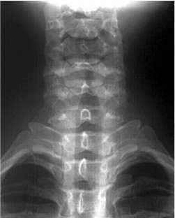
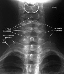
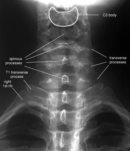
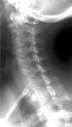
Normal AP (anterior-posterior) view of the cervical spine.
Usually
obtain three views, PA LAT and Ontontoid. Must see all 7 C-vertebrae (top of
T1). If C7 cannot be visualized,
get a swimmers view, if still not, a CT should be considered. Other plain film
views include oblique, flexion, and extension.
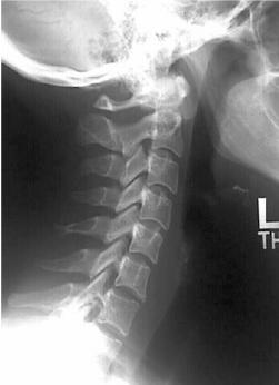

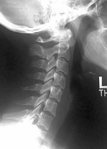
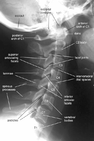
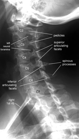
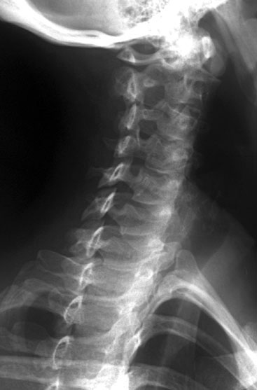
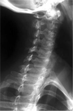
Normal LAT (lateral) view of the cervical spine.
Prevertebral
soft tissues should not measure >7mm at level of C3 and C4, and should not be
>20mm at the level of C6 ( not as reliable)
(In
children, 2/3 width of the C2 Vertebral body at the level of C3 and C4 and not
>14mm at the level of C6.)
If C2-
C6 soft tissues measure 7-10mm, consider further imaging.
If > 10mm definitely need
additional imaging if reason not apparent on plain film.
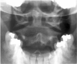
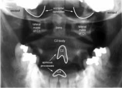
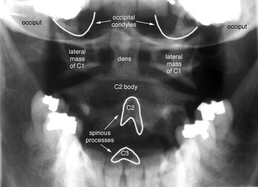
Normal Odontoid views
Flexion/Extension
– The posterior cortical margin of each cervical vertebral body may be offset
by as much as 3mm.
Note:
failure to visualize 7
cervical vertebrae is the most common error made in the
radiographic assessment of cervical spine injury.
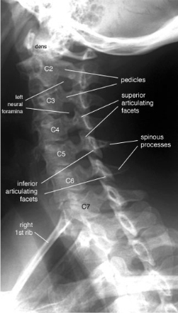
Normal
Left Anterior Oblique (LAO) position, allows visualization of the
Left-sided foramina.
Note: With
the head turned to the right (Left Anterior Oblique position), allows
visualization of the Left-sided foramina. The foramina to the left side of the
vertebral bodies will always be the left neural foramina.
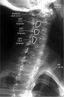
Normal Right Anterior Oblique (RAO) position, allows visualization of the
Right-sided foramina.
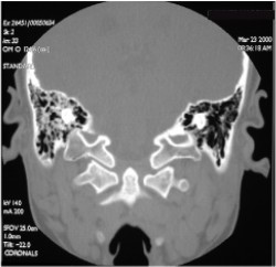
CT scan of T-bone and C1-C2
This section written by:
LCDR Ron Boucher, MC, USN
LT Hugh McSwain, MC, USN
With some assistance from:
CDR Michael Puckett, MC, USN
ENS Robert Post, MC, USNR
|