|
Thyroid Gland
It is shaped like a butterfly, and lies in the anterior part of the neck,
below the larynx. It consists of two lobes, one on each side of the upper trachea,
connected by a strip of tissue called the isthmus. The thyroid secretes the
iodine containing hormone THYROXIN, which controls the rate of cell metabolism.
Examine the Neck
-
Inspect the neck for masses or asymmetry.
- Evaluate range of motion and palpate for midline position of the trachea.
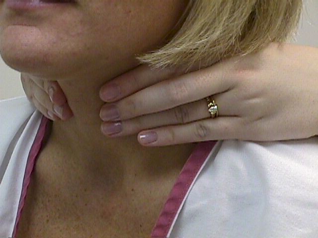
- Inspect and palpate the thyroid gland. It should be smooth,
symmetrical, and not enlarged.
- Inspect and palpate for lymph notes.
- If a node is enlarged or tender look for a source in the area that it drains. Tender nodes suggest inflammation; hard or fixed nodes suggest malignancy. Is it a node, muscle or artery: Remember you should be able to roll a node in two directions —up and down, and side to side — a muscle or artery will not pass this test.
- Check for nuchal rigidity — touch chin to sternum. Pain is a sign of meningeal irritation.

Source:
Operational Medicine 2001, Health
Care in Military Settings, NAVMED P-5139, May 1, 2001, Bureau
of Medicine and Surgery, Department of the Navy, 2300 E Street NW, Washington,
D.C., 20372-5300
|

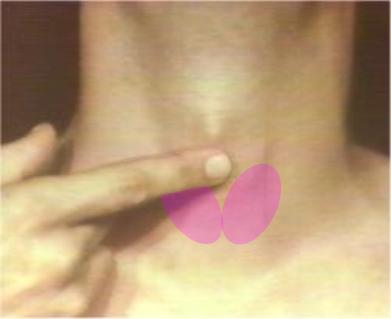
The index finger is on the cricothyroid
membrane
(below). The Thyroid
cartilage (Adam's Apple) is above
the finger, and the Cricoid cartilage
is
below the finger.

The two soft lobes of the thyroid gland,
fused in the
midline below the cricoid
cartilage, rise up on either side of the
trachea.
|