Hospital Corpsman Sickcall Screener's Handbook
BUMEDINST 6550:9A
Naval Hospital Great Lakes
1999
Dermatology
-
FUNCTIONS OF THE SKIN
-
Protection: a barrier against the unfriendly environment-keeping us in and the world out
-
Heat Regulation: The body loses heat by evaporation of sweat and by increased blood flow to the skin.
-
Sensory Perception: Fine touch, pressure, temperature, and pain.
-
LAYERS OF THE SKIN
-
Epidermis: provides the major part of the barrier
-
Dermis: contains blood vessels, provides support and nutrition for the epidermis, and is home to the nerves, sweat glands, hair follicles, and sebaceous glands.
-
Subcutaneous Fat Layer: provides insulation from cold and injury.
-
SKIN APPENDAGES
-
Sweat Glands: heat regulation and water and salt excretion.
-
Sebaceous Glands: found next to the hair follicles. They produce sebum which lubricates the skin and in larger quantities causes acne.
-
Hair: cosmetic importance.
-
Nails: protect the finger tips.
-
SKIN LESIONS AND CLINICAL DIAGNOSIS
-
The first and most importance step is to characterize the
appearance of each skin lesion:
-
Distribution on the body-localized or generalized over the body
-
Arrangement-grouped or isolated
-
Configuration-linear, annular (ring shaped), irregular (no pattern)
-
Primary and Secondary Lesions:
Primary Lesions are the first to appear on the initial presentation. Then the patient begins scratching or treats them, or they become infected. Over time the primary lesions become obliterated by the secondary lesions.
Primary Lesions:
-
Macule-a flat small (1cm) localized change in the color of the skin (a freckle,1st degree burn)
Two types: Erythema-redness due to capillary dilation, they blanch with pressure
-
Purpura-(purple-ish), do not blanch, they are deposits of blood.
-
Petechiae: very small (2mm) purpura.
-
Ecchymosis: large purpura
-
Papule: small (1cm) solid elevated lesion.
-
Plaque: a large papule.
-
Wheal: (hives/urticaria) a temporary edematous elevation usually with erythema.
-
Vesicles: small(1cm) fluid filled lesion (blister). Example: herpes, chicken pox.
-
Bulla: a large vesicle
-
Pustule: a pus filled vesicle (acne)
-
Furuncle-large pustule, deeper involvement
-
Carbuncle-several furuncles together
-
Abcess-a deep collection of pus
-
Comedo: a plug of sebum and bacteria in the hair follicle causing acne.
Secondary Lesions:
-
Scales: spontaneous shedding of the outer layer of the skin as in dandruff.
-
Crusts: an accumulation of dried fluid (serum or pus) on the skin surface. Usually the result of the rupture of a vesicle or pustule, as seen in the honey colored crusts of
impetigo.
-
Excoriations: loss of skin due to scratching.
-
Erosion: superficial loss of epidermis.
-
Lischenifiication: a thickening of the skin due to prolonged scratching. Hallmark of eczema.
-
History: Questions to Ask
-
What is the problem you are having with your skin?
-
How long have you had it? Acute, chronic, or recurrent?
-
What did the rash look like when it first started?
-
Does it itch?
-
How have you treated it?
-
Examination of the Skin: Examine the patient in good light, with exposure of the entire body.
There are six signs to identify:
-
Type of lesion: macule, papule, vesicle, etc.
-
Distribution: Location on the body-local or generalized.
-
Arrangement: isolated, grouped.
-
Shape of Lesion: linear, annular
-
Color: Red or purple, does it blanch?
-
Palpation of lesion: soft, firm, hard, moist, or dry?
-
Laboratory Aids:
-
Gram stains for bacteria
-
KOH for fungi or yeast.
-
If vesicular, take a glass slide and obtain a direct smear of the
base for giant cells as in Herpes.
-
Clinical Dermatological Problems:
-
Acne: Disease of sebaceous glands with onset of puberty. Comedos, pustules and erythematous papules on the face, chest, or back.
S: "Pimples" or "Zit"
O: Comedos (blackheads) and pustules on the face, chest, or back which may result in pitted scars.
A: Acne
P: Keep hands off the face and avoid squeezing lesions. Wash face BID.
-
Benzoyl Peroxide 5% applied once or twice daily
-
Retin A applied once daily
-
Tetracycline 250mg, two BID
-
Erythromycin 250mg, two BID
-
Folliculitis/Furuncles/Carbuncles:
Folliculitis is a localized infection of a hair follicle. Furuncle is a large deep follicular infection. Carbuncle is a large coalescence (joining together) of furuncles with several draining points usually found on the neck, back, or thighs.
S: Skin is painful, red, and swollen
O: Lesions vary in size, very tender and erythematous. Initially they are firm, but centers become fluctuant (movable and compressible). Regional lymphadenitis (inflamed lymph nodes may be present.)
A: Folliculitis, Furuncle, or Carbuncle
P: Folliculitis: Clean with soap and water, apply hot packs for 20 minutes QID
-
|
 |
|
Military
Medical CDs
Textbooks, Manuals, Instructions,
Videos
On-line Resources
www.brooksidepress.org |
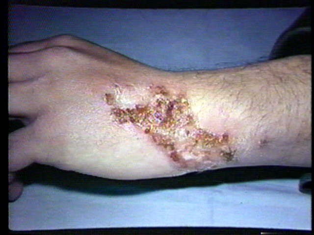
Impetigo:
An infection of the superficial layers of the skin caused by strep or staph bacteria.
S: A spreading of rash or sores
O: Honey colored crusted lesions usually of the face, with a erythematous base.
A: Impetigo
P: Dicloxacillin, or Erythromycin, or
Velosef 500mg QID for 10 days.
-
Cellulitis:
This is a deep infection of the skin caused by strep and staph. Patients with cellulitis of leg often have a preexisting lesion that acts as a portal of entry for the bacteria. Always check between the toes because tinea pedis may provide the portal of entry.
S: Patient may feel ill, usually have a fever. Has a large area of erythema that is swollen and painful.
O: Lesions that is red, warm, swollen, and tender. Lymph nodes tender and enlarged.
A: Cellulitis
P: Warm soaks, bed rest, keep part elevated
Dicloxacillin, Erythromycin, or
Velosef 500mg QID for 10 days
If not improved may need IV antibiotics
Facial cellulitis, common around the eye, requires hospitalization and IV antibiotics.
Refer to MD/PA
-
Pityriasis Rosea:
A self-limited mild, scaly, erythematous skin eruption occurring primarily in adolescents and young adults, lasting about 5 to 8 weeks.
S: A fine scaly rash, mild itching, but patient feels well
O: Oval papules and plaques with a delicate scar near the border of the lesions. Preceded by a "Herald Patch" Distributed generally, following the cleavage lines of the trunk-a pattern likened to a Christmas tree.
A: Pityriasis Rosea
P: No treatment is usually required
Test for syphilis with an RPR
-
Psoriasis: (over production of epidermis)
Psoriasis is a chronic disease characterized by over production of new skin. Instead of taking 19 days to replicate it takes only 1.5 days causing the skin to thicken forming the classic silver scale. Lesions have an irregular shape with a sharp border and a red base topped by a silver scale. The scalp, elbows, knees, groin, and feet are more commonly involved.
S: Itching may be mild to severe
O: Erythematous plaques covered with silvery scales. Pitted nails in 50% of patients
A: Psoriasis
P: Sunbathing or ultraviolet light treatments
Coal Tar compounds
Westcort Cream TID
-
Tinea Infection: (fungal)
Infection of the skin causing scaling, pruritis, and a red lesion with an elevated boarder. The most important lab test in the diagnosis of tinea is the potassium hydroxide or
KOH preparation.. The KOH dissolves normal cellwalls leaving the fungal cells visible, appearing as hyphae and spores when viewed under low power.
-
Tinea Cruris: an infection of the groin referred to as jock itch"
S: Burning, itching sensation in the groin
O: Lesions with scaling and an erythematous base and an elevated border. Lab-skin scrapings from the leading edge of the lesion show typical hyphae when prepare with KOH.
A: Tinea Cruris
P: Wear loose-fitting cotton underwear
Clotrimozole (Lotrimin, Mycelex) or Econazole (Spectozole) or Miconazole (Monistat
Derm) or Tolnaftate (Tinactin,
Pitrex). All of these are applied BID.
-
Tinea Pedis: Infection of the feet, the most common area affected. 2 types:
-
Interdigital-macerated scaling process between the toes
-
Vesiculopustular-vesicles and pustules on the instep-suspect tinea. A KOH
PREP taken from the underside of the roof of the vesicle or pustule will reveal fungal hyphae.
Treatment: same as for tinea cruruis.
-
Tinea Versicolor (varied color): Lesions are usually found on the trunk and upper arms and may vary in color from white to pink to tan, Usually asymptomatic. Diagnosis by
KOH.
Treatment: Selsun shampoo: allow to dry on skin overnight, and showered off in AM.
Repeated for 3 days. Note: It takes months for skin color to return to normal.
-
Seborrhea: (over production of sebum)
A chronic, superficial, inflammatory process with erythema and scaling affecting hairy regions of the body, especially the scalp, eyebrows, and face.
S: A scaly, pruritic rash on the scalp, eyebrows, and face
O: Dry to oily yellowish scales with erythema, secondary infection frequently present.
A: Seborrhea
P: Scalp: Shampoos with sulfur, coal, tar, or selenium
sulfide, rub into scalp, rinse off in 10 minutes. Topical steroids like Hydrocortisone Cream 1%, apply TID.
-
Eczema:
Eczema is a descriptive term only, not a specific disease. Under eczema are grouped skin problems that have eczematous inflammation consisting of redness, scaling, and vesicles and always itch. If it doesn't itch, don't consider eczema. If left alone eczema would resolve spontaneously, however with itching comes the scratching and irritation and thus develops the disease. Acute eczema itches intensely. Patients scratch the eruption even while sleeping. A hot shower temporarily relieves itching because the pain produced by hot water is better tolerated than the sensation of itching, heat aggravates acute eczema.
There are two stages to this problem and each has specific looking lesions.
-
Contact Dermatitis (Eczema)
This is an inflammatory response to a substance that has come into contact with the skin. There are two types:
-
Irritant contact dermatitis: has a direct toxic effect on the skin. Includes acids, alkalis, solvents and detergents.
-
Allergic contact dermatitis: triggers an immune response that causes tissue inflammation. Includes metals, plants (poison ivy), and medicines.
S: Itching, stinging, or burning at the site of contact. Erythema, vesicles, open weeping lesions.
O: Erythema, edema, vesicles, bullae, or weeping lesions may be present. The area is usually defined.
A: Contact Dermatitis
P: Antihistamines for pruritis:
Topical steroids for inflammation:
-
Hydrocortisone 1% or Westcort, or Aristocort creams, applied 3 to 4 times daily.
-
Burrow’s solution
(Domeborrows)-wet dressings dry weeping lesions.
-
Oral Steroids-for more severe cases esp. if due to poison ivy.
-
Watch for signs of secondary infection.
-
Lubricating oils and creams are helpful.
-
Atopic Dermatitis:
This is a chronic, pruritic, eczematous (redness, scaling, vesicles.) condition of the skin that is genetically determined and associated with a personal or family history of atopic disease (asthma, allergic rhinitis, dermatitis). Pruritis is the most distressing and prominent symptom. Lichenification is the clinical hallmark of atopic dermatitis. Secondary infection is common. In adults distribution includes the neck, face, upper chest, and the antecubital (anterior flexor surface of the elbow) and the popliteal fossae (back of the knees). First priority in treatment is stop the scratching.
S: Pruritis, scaling, dry, thickened skin
O: Red, weeping, and crusted lesions, lichenification, pruritis, usually found on the face, neck, and extremities. May have infection of excoriated (scratched) areas.
A: Atopic Dermatitis
P: Same treatment as for contact dermatitis.
-
Urticaria (Hives): An immunologic response to an allergenic stimuli as with drugs and foods, or a response to physical stimuli as with cold, pressure, sunlight, or rubbing/stoking of the skin (Dermographism). Characterized by a generalized distribution of wheals (hives), itching, and erythema. Lesions vary in shape from round or oval to confluent. There may be involvement of the lips, toungue, or eyelids. Hives may last a few hours to a few weeks. If allergic response is severe it may lead to anaphylactic shock, respiratory distress, and sudden death.
S: Hives, Itching
O: Pruritis, raised wheal-like skin lesions on any area of the body, erythema.
A: Urticaria
P: If possible remove the cause.
Antihistamines:
During the day: Seldane one tab PO BID, little sedation
Evening: Atarax 10 to 25mg every 6 hours, if nor responsive to
Atarax try
Diphenhydramine (Benadryl) 50mg q 6 hours.
In severe cases (anaphylactic shock):
Epinephrine 1:1000 .3 to .5ml IM
Benadryl 50mg IM
Refer to MD
-
Scabies:
A parasitic infection of the skin. The female burrows into the skin, deposits eggs which hatch in 1-2 weeks. Areas of involvement include the fingers, wrist, elbows, waist, and penis. Burrows. Nodules, or vesicles may be visible. Diagnosis is confirmed by scraping the lesion and adding mineral oil to the slide and identifying the parasite or its eggs under low power.
S: Itching, worse at night. Small red bumps on the sides and web spaces of the fingers and wrists.
O: Pruritic, may see burrow, nodules on the penis, usually involving the fingers and wrists. Scabies identified by scraping.
A: Scabies
P: Kwell Cream. Apply to all skin surfaces below the neck and wash off in 8 to 12 hours. Reapply to hands if they are washed. It is normal to continue to itch for days or weeks after treatment-further use of Kwell may cause dermatitis and worsen itching. Need to treat family or roommates. Repeat application in one week if scabies are identified.
-
Pediculosis Pubis (pubic or crab louse):
An infestation with lice that is transmitted by close contact. They live on, rather than in, the body, feeding 5 times a day. They are active and can travel quickly and survive for a week when separated from a host. Lice and eggs (nits) can be found cemented to the bases of hair shafts close to the skin.
S: Itching of the affected area
O: Lice and/or nits seen in pubic area
A: Pediculosis Pubis (crabs)
P: Kwell Cream or shampoo. Check the pharmacy section for proper use.
-
Warts (Verrucae):
The common wart or verrucae is flesh colored, dome shaped, firm papule that has a corrugated surface. It interrupts the normal skin lines and is studded with black dots which are thrombosed capillaries (a useful diagnostic sign-easily seen after paring or slicing away the surface of the wart). The normal skin line return when the wart is gone-a good sign of cure. The hands are the most common site but warts may be found on any skin surface. Warts are caused by the human papillomavirus (HPV). On the feet they are called planar warts. They are flat because of the constant pressure. Flat warts are usually found on the forehead. Subungal and periungal warts-found under and around the nails, are resistant to treatment because much of the wart may be submerged under the nail.
S: Wart-anywhere on the body
O: Flesh colored firm growth, shape, size and appearance may vary. Normal skin lines interrupted.
A: Warts (Verrucae)
P: Treatment varies:
-
Cryotherapy (freezing) with liquid nitrogen (Do not freeze warts on the feet)
-
Salicylic acid plasters (Mediplast), cut to the size of the wart and apply.
Follow instructions found in the pharmacy section.
-
Retin A cream applied at bedtime over the entire area involved will usually clear flat warts to the forehead.
Note: Genital warts and mulluscum contagiosum are covered in the STD section.
-
Skin Cancers:
Prime risk factor is intense sun exposure-use sun screens especially if fair skinned. Patients should be asked about any new, slowly growing lesions that are flesh colored, any history of bleeding or ulceration of lesions. Areas of maximum solar exposure are at risk. In malignant melanoma-look for any
pigmented lesion that has an irregular boarder, variations in color, especially blue.
|
|
Approved for public release;
Distribution is unlimited.
The listing of any non-Federal product in this CD is not an endorsement of the
product itself, but simply an acknowledgement of the source.
Bureau of Medicine and Surgery
Department of the Navy
2300 E Street NW
Washington, D.C
20372-5300 |
Operational Medicine
Health Care in Military Settings
CAPT Michael John Hughey, MC, USNR
NAVMED P-5139
January 1, 2001 |
United States Special Operations
Command
7701 Tampa Point Blvd.
MacDill AFB, Florida
33621-5323 |
*This web version is provided by
The Brookside Associates Medical Education Division. It contains
original contents from the official US Navy NAVMED P-5139, but has been
reformatted for web access and includes advertising and links that were not
present in the original version. This web version has not been approved by the
Department of the Navy or the Department of Defense. The presence of any
advertising on these pages does not constitute an endorsement of that product or
service by either the US Department of Defense or the Brookside Associates. The
Brookside Associates is a private organization, not affiliated with the United
States Department of Defense.
Contact Us · · Other
Brookside Products

|
|
Operational Medicine 2001
Contents
|
|
 |
|
FMST Student Manual Multimedia CD
30 Operational Medicine Textbooks/Manuals
30 Operational Medicine Videos
"Just in Time" Initial and Refresher Training
Durable Field-Deployable Storage Case |
|