1-1. THE SPECIAL SENSES
a. The human body is continuously bombarded by all kinds of stimuli. Some of these stimuli are received by sensory receptors distributed throughout the entire body. Other stimuli are received by highly complex receptor organs. These are referred to as the special senses.
b. From each special sense organ, information is sent to the brain through specific cranial nerves. When the information reaches the specific area of the brain’s cerebral cortex, it is perceived at the conscious level as sight, sound, smell, taste, and balance. These special senses allow us to detect changes in our environment, providing information necessary for homeostasis.
c. The Special Senses are:
(1) Sight.
(a) The receptor organ is the eye.
(b) The stimulus is light rays.
(2) Hearing.
(a) The receptor organ is the ear (cochlea).
(b) The stimulus is sound waves.
(3) Smell.
(a) The receptor organ is the nose (olfactory hair cells).
(b) The stimulus is airborne molecules.
(4) Taste.
(a) The receptor organs are the taste buds in the mouth.
(b) The stimulus is fluid-borne molecules.
(5) Balance (equilibrium).
(a) The receptor organ is the ear (membranous labyrinth).
(b) The stimulus is gravitational forces.
1-2. SMELL
Located in the upper recesses of the nasal chambers is a special layer of tissue called olfactory epithelium. Within the olfactory epithelium are special hair cells (chemoreceptors) that react to airborne molecules. Information received by the hair cells is transmitted from the olfactory nerves to the olfactory bulbs and along the olfactory tract to the brain, where it is interpreted as the sensation of smell.
1-3. TASTE
Located on the tongue and back of the mouth are sensory receptors called taste buds. Special hair cells in the taste buds are chemoreceptors that react to molecules of material taken into the mouth (food, liquids, and so forth.). Information received by the hair cells is transmitted to the brain, where it is interpreted as the sense of taste.
1-4. VISION
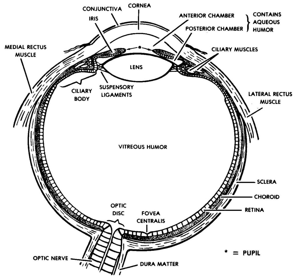
The eye (figure 1-1) is the special sense organ responsible for vision. Rays of light (reflected from an object) pass through the cornea, aqueous humor, pupil, lens, and vitreous humor to stimulate the receptor tissue (rods and cones) in the retina. The resulting nerve impulses travel to the brain, where they are interpreted as sight.

1-5. HEARING
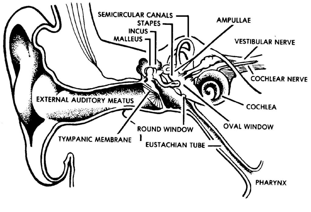
The human ear (figure 1-2) serves two major sensory functions–hearing and equilibrium.
a. Sound stimuli travel as airborne waves, which are collected by the external ear. The airborne waves pass through the external auditory meatus (ear canal) to the tympanic membrane, which separates the external and middle ear.
b. The physical vibration of the airborne waves is converted to mechanical vibration by the tympanic membrane and the ossicles. The ossicles (malleus, incus, and stapes) articulate with both the tympanic membrane and the oval window, which opens into the vestibule of the inner ear.
c. When the ossicles are set into mechanical vibration, the stapes acts as a plunger against the oval window, imparting pressure pulses to the fluid (perilymph) of the inner ear.

d. Fluid vibrations of the perilymph are converted to nerve impulses when the hair cell receptors within the cochlea are stimulated by the fluid vibrations. The nerve impulses are carried to the brain where they are interpreted as sound.
1-6. EQUILIBRIUM
a. Equilibrium is the state of balance of the body. Through a variety of sensory inputs and postural reflexes, the body can be maintained in a desired posture. All of the various sensory inputs related to the maintenance of equilibrium and posture are integrated within the brain as “body sense.” The internal ear provides one of the input systems for general body sense.
b. A primary sensory input for equilibrium consists of gravitational forces. Gravitational forces are of two types: static, when the body is standing still, and kinetic, when the body is in motion. Kinetic motion may be in a straight line (linear), or in an angular direction (curvilinear).
c. The fluid-filled membranous labyrinth of the inner ear has two sac-like structures called the sacculus and the utriculus. On the wall of each sac is a collection of special hair cells, which serve as receptors for static and linear kinetic gravitational forces.
d. Associated with the utriculus are three tubular structures called the semicircular canals. Two of the semicircular canals are vertically oriented and the third is essentially horizontal. All three semicircular canals are oriented at right angles to each other.
e. Each semicircular canal ends with an enlarged area where it opens into the utriculus. This area is called the ampulla. On the wall of each ampulla, at a right angle to the axis of the canal, is a little ridge of hair cells. The hair cells bend in directional response to the kinetic gravitational forces initiated by movement of the head.
f. All the information from the hair cells of the sacculus, utriculus, and ampullae is transmitted to the brain by the vestibular nerve. The vestibular and auditory nerves are contained within the same fibrous sheath from the inner ear to the brain. Within the brain, the two nerves split into different pathways.
