Pediatric Otitis Media
|
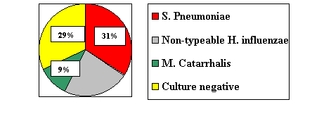
Otitis media is a common childhood illness whose pathogenesis appears primarily from eustachian tube dysfunction, which may be due to adenoiditis, allergy, chemical irritation (second hand smoke), upper respiratory illnesses, or congenital anomalies. Eustachian tube dysfunction causes changes in pressure (high negative pressure) in the tiny middle ear cavity, resulting in a sterile transudate which subsequently becomes contaminated with infected nasopharyngeal contents by aspirations or insufflation during crying and nose blowing. Organisms include Streptococcus pneumoniae, Hemophilus influenzae, group A streptococcus, and Moraxella catarrhalis.
Document the presence or absence of otalgia, aural discharge, fever, nausea, vomiting, appetite, fluid intake, URI symptoms, exposure to cigarette smoke, attendance at day care, past otitis history, present otitis history, past and present medication history, drug allergies, irritability, and other illness in the family. Be attentive to the documentation of past incidences of otitis, including the response to specific antibiotics and the results of follow up examinations. On physical examination, make note of the child's appearance (visual alertness, interest in engaging with examiner, consolability). Document the presence or absence of conjunctivitis, pharyngitis, tonsillitis, adenitis, meningeal signs, pain on moving the pinna, and aural discharge. Otitis media is defined by an abnormal tympanic membrane (TM). Describe four aspects of the tympanic membranes (PCTM).
Differentiating normal from abnormal A normal TM will be in a neutral position with a pearly gray color, with easily visible landmarks (translucent), and with good mobility to both positive and negative pressure. A TM that has fluid or pus behind it will be dull or full appearing, with an abnormal color (yellow, red, or occasionally blue), without landmarks, and without normal mobility. Document the presence or absence of fluid level or air bubbles. Look for perforation, tympanostomy tubes, cleft palate, or other deformity. Audiometry has limited value in the diagnosis of AOM, but tympanometry is helpful in the detection of middle ear effusion, especially in cases where the diagnosis is uncertain clinically. Audiometry is helpful in documenting hearing loss associated with otitis media externa (OME) or chronic otitis media (COM). Nasal and pharyngeal cultures show poor correlation with results of cultures taken at the time of myringotomy. Differential diagnosis and Referral criteria The differential diagnosis of ear pain includes pharyngitis, dental disease, temporal mandibular joint (TMJ) disease, and external otitis. The following patients should be referred to a pediatrician:
Treatment considerations and medication options The treatment of AOM (in children older than 8 weeks) is becoming controversial. Since over 25 percent of all cases of AOM are culture negative (presumed viral) and another 25 percent of cases of culture positive AOM will resolve spontaneously and have no benefit from antibiotic therapy, many now advocate observation for 2-3 days with supportive treatment (analgesics and nasal decongestants). If the child does not improve by 72 hours, then antibiotics should be instituted. Although this philosophy has not caught on widely in the United States, one must remember that the cause of the emergence of antibiotic resistance can be directly attributed the over treatment of otitis media and that the likelihood of aural or intracranial complications are exceedingly rare in Europe, where may hold this "72 hour" philosophy. Remember that it is very common for children to have URIs and have serous fluid in the ears. If antibiotic treatment is warranted, the following oral antibiotics are recommended for 10 days:
*Indicates first line therapy. Doses are located in the Pediatric Formulary Section of this manual. Treatment vs. Compliance failure There is recent evidence that approximately half of patients with acute otitis media may have a concurrent viral infection in the middle ear. This may explain why some cases of otitis media do not respond to antibiotics. If a child with otitis does not respond to treatment, there are two possibilities: treatment failure or compliance failure. If the child has received the medication as prescribed, (be absolutely certain of this before ascribing the lack of response to treatment failure) and still has an acute suppurative otitis media, then the infection is either viral or due to a resistant bacteria. Choose a different antibiotic and continue therapy, or better yet, obtain a tympanocentesis for culture (call ENT). Penicillin-resistant pneumococcus represents one of the biggest health care challenges. Up to 30-40 percent of isolates are Amoxicillin resistant. High-dose Amoxicillin (80mg/kg/day) is still recommended. If after 10 days of an appropriate antibiotic treatment, the child is without pain, afebrile, eating well, and sleeping well, but with a persistent effusion, observe for another 10 to 12 weeks (it may take the Eustachian tube 8-12 weeks to recover fully). If effusion persists after this period, refer to an ENT specialist. OME should, at the most, be treated with one course of a beta-lactam stable antibiotic for 10-14 days. Further treatment is futile and expensive. In some studies, oral steroids have shown to be beneficial, but are of limited long-term value. Use of antihistamines and decongestants in the treatment of AOM or OME is not efficacious. One caveat to this is those patients with documented seasonal or perennial allergic rhinitis. Nasal steroids have also been recently advocated for OME, but its role has yet to be defined. Final notes Consider referred to ENT for surgical management if a child has more than 3 bouts of AOM in 6 months (separate cases) or 4 in a year or any aural or intracranial complications. OME present for longer than 3-4 months should also be referred. If speech delay is present, initiate a speech consult. Reviewed by CDR Wendy Bailey, MC, USN, Pediatric Specialty Leader, Department of Pediatrics, Naval Medical Center San Diego, San Diego, CA (1999). |
Preface · Administrative Section · Clinical Section
The
General Medical Officer Manual , NAVMEDPUB 5134, January 1, 2000
Bureau
of Medicine and Surgery, Department of the Navy, 2300 E Street NW, Washington, D.C.,
20372-5300
This web version of The General Medical Officer Manual, NAVMEDPUB 5134 is provided by The Brookside Associates Medical Education Division. It contains original contents from the official US Navy version, but has been reformatted for web access and includes advertising and links that were not present in the original version. This web version has not been approved by the Department of the Navy or the Department of Defense. The presence of any advertising on these pages does not constitute an endorsement of that product or service by either the Department of Defense or the Brookside Associates. The Brookside Associates is a private organization, not affiliated with the United States Department of Defense. All material in this version is unclassified. This formatting © 2006 Medical Education Division, Brookside Associates, Ltd. All rights reserved.
Home · Textbooks and Manuals · Videos · Lectures · Distance Learning · Training · Operational Safety · Search
This website is dedicated to the development and dissemination of medical information that may be useful to those who practice Operational Medicine. This website is privately-held and not connected to any governmental agency. The views expressed here are those of the authors, and unless otherwise noted, do not necessarily reflect the views of
the Brookside Associates, Ltd., any governmental or private organizations. All writings, discussions, and publications on this website are unclassified.
© 2006 Medical Education Division, Brookside Associates, Ltd. All rights reserved
Other Brookside Products
