TABLE OF CONTENTS
INTRODUCTION
1 ANATOMY AND PHYSIOLOGY OF THE NERVOUS SYSTEM
Exercises
2 PHYSICAL ASSESSMENT OF THE NERVOUS SYSTEM
Exercises
3 CENTRAL NERVOUS SYSTEM DISEASES AND DISORDERS
Section I. Diseases of the Central Nervous System
Section II. Disorders of the Central Nervous System
Exercises
4 SEIZURES
Exercises
5 CENTRAL NERVOUS SYSTEM TRAUMA
Section I. Head Injury
Section II. Spinal Cord Injury
Section III. Immobilization Techniques for Spinal
Cord Injury
Section IV. Management of Spinal Cord Injury
Exercises
---------------------------------------------------
LESSON 1
ANATOMY AND PHYSIOLOGY OF THE NERVOUS SYSTEM
1-1. INTRODUCTION
The nervous system facilitates contact of the
individual with his external and internal environments and aids in
appropriate responses to these constantly changing environments. A
general knowledge of the anatomy of the nervous system and an
understanding of its physiology will help you to recognize and treat
injuries and diseases of the nervous system.
1-2. ROLE OF THE NERVOUS SYSTEM
a. The nervous system has three general functions that
it performs in the role of the body's control center and communication
network. The functions are:
(1) The nervous system is able to sense change both
inside the body and change in the environment surrounding the body.
(2) The nervous system is able to interpret these
changes.
(3) The nervous system causes the body to react to
these changes by either muscular contraction or glandular secretion.
b. Homeostasis is a good example of the nervous system
sensing change, interpreting change, and adjusting to change. (In
homeostasis, the equilibrium of factors such as temperature, blood
pressure, and chemicals are kept in relative balance.) In the case of
homeostasis, the nervous system and the endocrine system operate
together to maintain equilibrium in the body.
1-3. ORGANIZATION OF THE NERVOUS SYSTEM
The nervous system has two main divisions: the central
nervous system (CNS) and the peripheral nervous system (PNS). The
central nervous system is composed of the brain and the spinal cord.
This system controls behavior. All body sensations are sent by
receptors to the central nervous system to be interpreted and acted
upon. All nerve impulses that stimulate muscles to contract and glands
to secrete substances get the message from the central nervous system.
The peripheral nervous system is composed of nerves. This system is a
pathway to and from internal organs. PNS serves as a pathway to the
brain for the five senses and helps humans adjust to the world around
them. Further subdivision of the peripheral nervous system will be
discussed later in this lesson.
1-4. CELL ORGANIZATION OF THE NERVOUS SYSTEM
Only two principal kinds of cells exist in the nervous
system: neurons and neuroglia. Neuroglia cells (also called glial
cells) act as connective tissue and function in the roles of support
and protection. Some of these cells twine around nerve cells or line
certain structures in the brain and spinal cord. Other neuroglia cells
bind nervous tissue to supporting structures and attach neurons to
their blood vessels. Other small neuroglia cells protect the central
nervous system from disease by surrounding invading microbes and
clearing away debris. Clinically, these cells are important because
they are a common source of tumors of the nervous system. Neuron cells
are nerve cells, the basic unit that carries out the work of the
nervous system. Impulses from one body part to another body part are
conducted by neurons.
1-5. COMPONENTS OF NEURONS
The neuron, the basic unit that carries out the work
of the nervous system, is a specialized conductor cell that receives
and transmits electrochemical nerve impulses. In other words, neurons
are nerve cells that conduct impulses from one body part to another
body part. Each neuron is made up of three distinct parts: the cell
body, dendrites, and an axon.
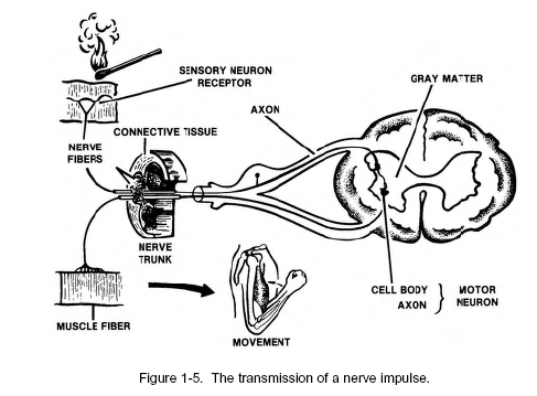
a. Cell Body, Dendrites, and Axon. The cell
body contains a nucleus or control center. Also, a neuron usually has
several highly branched, thick extensions of cytoplasm called
dendrites. The exception is a sensory neuron that has a single, long
dendrite instead of many dendrites. At the other extreme are motor
neurons, each of which has many thick "tree-like" dendrites. The
dendrite's function is to carry a nerve impulse toward the cell body.
An axon is a long, thin process that carries impulses away from the
cell body to another neuron or tissue. There is usually only one axon
per neuron. Axons vary in length and diameter and are "jelly-like" in
appearance.
b. Myelin Sheath (Schwann Cells). The myelin
sheath is a white segmented covering made up of Schwann cells. The
covering is around axons and dendrites of many peripheral neurons.
This covering wraps around the entire axon in "jelly-roll" fashion,
except at the point of termination and at the nodes of Ranvier. (The
nodes of Ranvier are intermittent constrictors along the myelin
sheath.) The myelin sheath is made up of a layer of protein, two
layers of lipids or fats, and one more layer of protein.
c. Neurilemma. The neurilemma is the nucleated
cytoplasmic layer of the Schwann cell. The neurilemma allows damaged
nerves to regenerate. Nerves in the brain and spinal cord DO NOT have
a neurilemma and, therefore, DO NOT recover when damaged.
From The Central
Nervous System


