NURSING CARE RELATED TO THE SENSORY AND
NEUROLOGICAL SYSTEMS
2-3
2-3. THE BRAIN
The human brain has three major subdivisions--the brainstem, the cerebellum, and the cerebrum.
a. The Brainstem. The brainstem (figure 2-1) is the basal portion of the brain. It is continuous with the spinal cord. Exiting from the sides of the brainstem are 12 pairs of nerves known as the cranial nerves. The brainstem is divided into 3 major portions.
(1) Forebrainstem (diencephalon), consisting of:
(a) Thalamus--a sensory relay station for impulses conveyed upward from the spinal cord.
(b) Hypothalamus--concerned with regulation of autonomic functions.
(2) Midbrainstem (mesencephalon) consisting of:
(a) Corpora quadrigemina--concerned with vision and hearing.
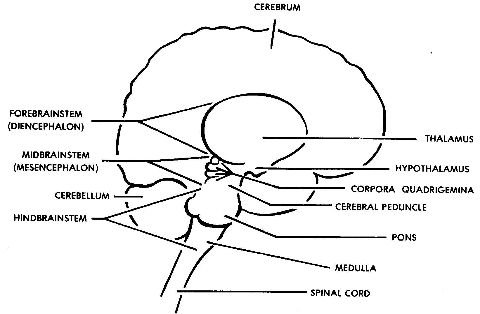
Figure 2-1. The brainstem.
(b) Cerebral peduncles--connecting the brainstem to the cerebrum.
(3) Hindbrainstem, consisting of:
(a) Pons--concerned with transmission of impulses between the left and right hemispheres of the cerebellum.
(b) Medulla--contains nerve centers for cardiac, vasomotor, and respiratory functions. Also influences sneezing, coughing, hiccoughing, and vomiting.
b. The Cerebellum. The cerebellum (figure 2-2) is a spherical mass of nervous tissue attached to and covering the hindbrainstem. It is attached to the brainstem by three pairs of cerebellar peduncles. The cerebellum consists of three lobes; the right hemisphere, left hemisphere, and a central portion called the vermis.
(1) The outer layer, or cortex, of the cerebellum is composed of "gray matter." Gray matter is actually the cell bodies of neurons. Many folds (gyri) and grooves (sulci) in the surface of the cortex give it a wrinkled appearance.
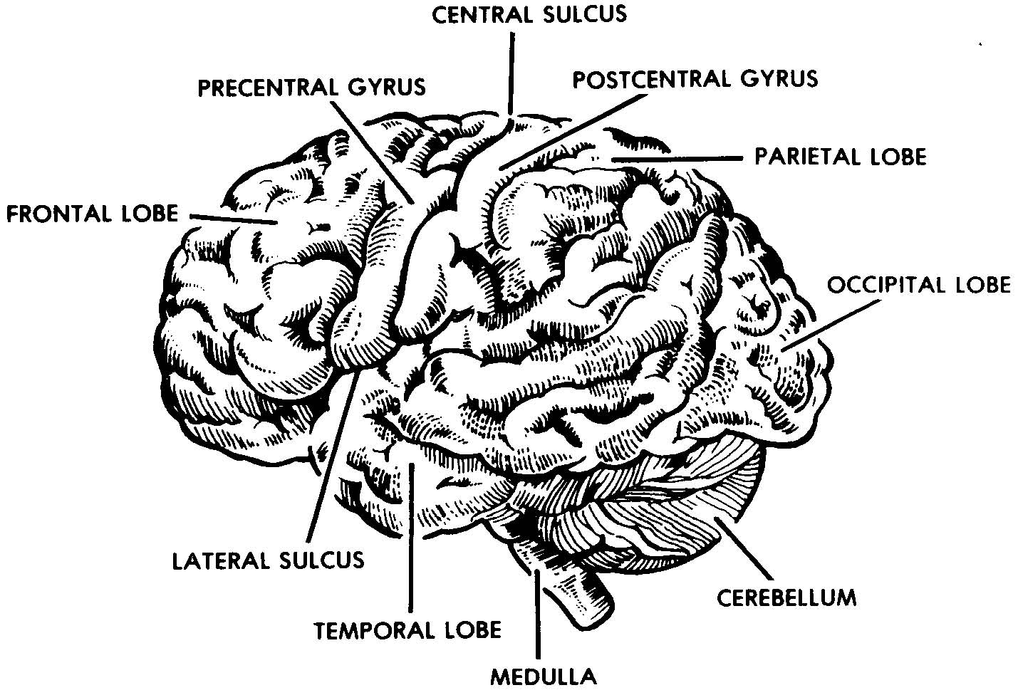
Figure 2-2. Human brain, lateral view.
(2) More centrally located within the cerebellum is the "white matter." White matter is actually the myelin covered processes of the neurons.
(3) The cerebellum is concerned with coordination of nerve impulses to the voluntary muscles and with the equilibrium of the body.
c. The Cerebrum. The cerebrum (figure 2-2) is the largest part of the brain. The outer layer of the cerebrum is called the cerebral cortex. It is composed of the cell bodies of neurons and is often referred to as the "gray matter." Many folds (gyri) and grooves (sulci) increase the surface area of this layer. Beneath the cortex lies the "white matter," actually the myelin covered processes of the neurons. The cerebrum is divided into right and left hemispheres by a longitudinal fissure. A band of nerve fibers called the corpus callosum connects
the hemispheres and provides for communication between them. Each cerebral hemisphere is further divided into lobes, which are named after the overlying cranial bones (frontal, parietal, temporal, and occipital).
(1) Frontal lobe.
(a) The frontal lobe is located beneath the frontal bones of the skull. It is divided from the parietal lobes by the central sulcus and from the temporal lobes by the lateral fissure.
(b) The functions of the frontal lobe include personality, behavior, intellectual functioning, creative thinking, morals, ethics, and level of consciousness.
(c) The precentral gyrus, within the frontal lobe and directly in front of the central sulcus, initiates and controls voluntary muscle movements (motor function). Impulses originating here travel along motor pathways through the spinal cord and stimulate skeletal muscles on the opposite side of the body. (This is important to remember when correlating clinical symptoms with associated cerebral damage.) The percentral gyrus also contains Broca's speech center, which is involved in the motor activities necessary for speech.
(2) Parietal lobes.
(a) The parietal lobes are located beneath the parietal bones of the skull. They are posterior to the central sulcus and superior to the lateral fissure.
(b) The functions of the parietal lobes include comprehension of written and spoken language, discrimination of fine touch, and stereogenesis. Sterogenesis is the ability to recognize, by touch alone, the size, shape, texture, and consistency of objects.
(c) The postcentral gyrus, within the parietal lobes and directly behind the central sulcus, controls and interprets sensations from the opposite side of the body. These sensations include pain, heat, cold, and pressure.
(3) Temporal lobes.
(a) The temporal lobes are located beneath the temporal bones of the skull and inferior to the lateral fissure.
(b) The functions of the temporal lobes include taste, smell, balance, and hearing. (The perception of sound as well as the interpretation of sound as words.)
(c) The area of the brain concerned with the comprehension of both written and spoken language (Wernicke's area) is located in both the parietal and temporal lobes.
(4) Occipital lobe.
(a) The occipital lobe is located beneath the occipital bone of the skull, at the posterior of the cerebrum behind the parietal lobes.
(b) The occipital lobe receives and interprets visual stimuli.