a. Tension Pneumothorax.
Tension pneumothorax is a condition in which air enters the pleural cavity outside the lung and becomes trapped. As more and more air becomes trapped, the increased pressure causes the lung in the affected pleural cavity to collapse. Tension pneumothorax can be caused by an open chest wound (Section II), but it can also result from a closed chest injury.
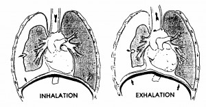
Figure 3-11 shows tension pneumothorax resulting from air that has escaped from a lung injured by floating rib segments.
Tension pneumothorax can also result from damage to the bronchi (air tubes) leading to the lungs. Air in the pleural cavity that results from disease rather than trauma to the lung is referred to as spontaneous pneumothorax. Complete collapse of the lung may be followed by tracheal deviation and mediastinal shift. Signs of tension pneumothorax include increased difficulty in breathing, shortness of breath, absent or diminished breath sounds on the effected side, subcutaneous emphysema, distended neck veins, bulging chest tissues, weak pulse, and cyanosis.

b. Hemothorax.
Hemothorax is a condition in which blood enters the pleural cavity outside the lung and becomes trapped.
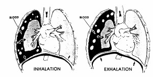
As more and more blood becomes trapped, the increased pressure causes the lung in the affected pleural cavity to collapse (figure 3-12). Hemothorax can be caused by any chest injury. It can result from lacerated blood vessels in the chest wall, lacerated major blood vessels within the chest, or laceration of the lung. Signs and symptoms of tension pneumothorax also apply to hemothorax. In addition, hemothorax may result in hypovolemic shock.
Tension pneumothorax and hemothorax may be present together. A hemothorax is more likely to cause significant hypovolemia before tension could be built up. The left and right lung spaces and the mediastinum can hold more than three liters of fluid (over half of the circulating blood volume).
NOTE: Figures 3-11 and 3-12 do not show a significant trachea deviation or mediastinal shift. As the pressure increases and the injured (right) lung collapses, the trachea and heart will be pushed more and more toward the casualty’s uninjured (left) side. The shift will compress the heart and the uninjured (left) lung.

