Shoulder Pain

The shoulder is a complex, unstable, and intricate joint that can be injured both through trauma and overuse. 

Shoulder pain is usually due to injury to one of three main groups of shoulder structures:

  • The muscles

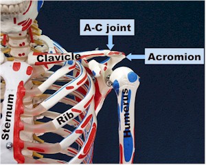
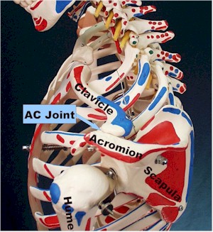
  • The bones and the AC joint (Acromio-clavicular joint)

  • The glenohumeral joint.  

Determine the mechanism of injury and the extent of disability.  A good history and physical will usually determine the cause of the pain and direct the care.

Differential diagnosis of shoulder pain:

Acute injuries 

  • Bones and AC joint

    • AC joint separation

    • Clavicle fracture

    • Humerus fracture- surgical neck, tuberosities

    • Scapular fracture – acromial process, coracoid process, body, spine

  • Glenohumeral joint

    • Dislocations, anterior and posterior

    • Subluxations (partial dislocations)

    • Labral tears

  • Muscles

    • Rotator cuff tears/strains

    • Biceps tear

    • Deltoid, trapezius strain

    • Pectoralis major tear                       

Chronic injuries

  • Bones and AC joint

    • AC joint capsulitis

  • Glenohumeral joint

    • Instability

    • Chronic labral tear

    • Degenerative joint disease

    • Frozen shoulder

  • Muscles

    • Rotator cuff impingement and degenerative tear

    • Tendonitis of other muscles

Referred pain

  • Causes include heart attack, cervical or brachial plexus nerve injury, cholecystitis, Pancoast tumor, pneumothorax, aortic aneurysm, axillary thrombosis.

History and physical

Always verify the distal pulses, capillary refill, and sensation.

The key to understanding shoulder pain is understanding the anatomy:

  • The shoulder has only one bony attachment to the chest – the clavicle.  

  • The shoulder has a wide range of motion because of the mobility of both the glenohumeral joint and the scapulothoracic articulation.  

  • The trapezius, deltoid, and pectoralis muscles are major superficial structures around the joint.  

  • Any of the surrounding muscles can have acute or chronic strain/tear.  

  • Pain with specific movements and tenderness in the muscle belly will confirm the diagnosis.

The bony structures and AC joint are usually injured through trauma:

  • A fracture can occur to any part of the bones.  

  • Palpation of the clavicle will elicit pain if fractured.  

  • Pain on gentle range of motion of the arm suggests humerus fracture.  

  • Scapular fractures are often hard to detect on physical.  

  • The Acromio-clavicular joint (AC joint) can either be damaged through trauma, as in a direct fall on the shoulder causing an AC separation, or through overuse , especially in weight lifters who get AC joint capsulitis and necrosis.

The glenohumeral joint is made of the glenoid fossa of the scapula, the humeral head, the joint capsule, and the labral cartilage, essentially the meniscus of the shoulder which helps hold the humeral head in place.  These structures are usually injured through trauma, especially with an anterior dislocation or subluxation.  

  • When the arm is forcibly abducted and externally rotated, the humeral head will partially (subluxation) or completely slide out of the joint anteriorly AND inferiorly, ripping joint capsule and possibly the labrum as it goes.  

  • Posterior dislocations are much more rare and come from direct blows to the outstretched arm forcing the humeral head posteriorly.  

  • The patient with a dislocated shoulder will hold their arm at their side and be unable to touch the opposite shoulder.  

  • A first time dislocator will need assistance to put the shoulder back in, the subluxer never came completely out so does not need relocated.  

  • Part of the anterior inferior glenoid rim may tear off as the humeral head slides off – this is called a bony Bankhart lesion.  

  • Labral tears will often have a painful clicking sensation.

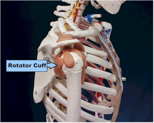
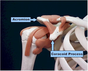
The rotator cuff muscles all originate off the scapula and wrap around the humerus, helping to hold the humerus in the glenoid fossa.  The subscapularis comes from the underside of the scapula and wraps around to attach anteriorly on the humerus; it internally rotates the arm.  The supraspinatous originates above the spine of the scapula, runs under the acromium and over the humerus to attach on the lateral humerus, abducting the arm.  It is the muscle that commonly gets impinged between the acromium and humerus and is usually the one torn in rotator cuff tears.  The infraspinatous and teres minor come from the lower scapula attaching to the posterior humerus.  Therefore, when they contract they externally rotate the humerus.  

The rotator cuff is rarely torn in young people without a dramatic injury such as when a baseball pitcher blows out his arm.  Older folks get degenerative tears.  

Testing of the rotator cuff muscles is done by testing internal rotation (subscap.),  external rotation (teres minor and infrasp.) and abduction with the thumbs down and arms parallel to the ground (supraspin.).  The impingement test is done by having the arm 90 degrees forward flexed parallel to the ground and then gently internally rotating.  Pain indicates a positive test.

When should x-rays be obtained?

  • Suspected fracture

  • Following reduction of a dislocation (some advocate x-rays prior to relocation)

  • Consider chest x-ray and c-spine x-ray if referred pain is possible

  • Persistent pain, night pain, unexplained weight loss – to look for bony tumor

What other studies should be considered?

  • An MRI can show a rotator cuff tear.  Adding contrast increases sensitivity for a labral tear, although arthroscopy is often necessary to detect labral tears

  • MRI of the C spine following x-rays if radiculopathy is suspected cause

  • Arthrogram can be used to rule out a complete tear of the rotator cuff

  • Other tests for referred pain if suspected as cause (e.g. enzymes, EKG for MI)

Treatment plan.

Treatment depends on the severity of the injury.  

  • Severely injured shoulders such as suspected fractures, separations, etc., should be placed in a sling and given pain medicines.  

  • Consider emergent referral for neurovascular compromise.  

  • Dislocated shoulders should be reduced.  

  • Less severe injuries will need less immobilization and can begin rehabilitation sooner.  

  • Use pain as a guide.  Specific lesions will be described in more detail later.

In general , follow the guidelines for treatment of acute injuries: PRICEMM

  • Protection:  Sling.  Patients with severe pain and disability such as in a suspected fracture, shoulder subluxation, or AC separation should be placed in a sling for comfort.

  • Relative rest.  Gentle pendulum exercises can usually be performed to help prevent a frozen shoulder

  • Ice 20 minutes three times a day

  • Elevate if possible

  • Medications – pain control as necessary

  • Modalities  - if available 

Rehabilitation begins with ranges of motion exercises.   Pendulum exercises where the patient leans over and gentle rotates the arm in a circular motion are usually the first activities started.  Gradually increase motion, then work on strength.  Rotator cuff strengthening exercises are useful in many conditions and are essential for treatment of impingement syndrome.  Endurance is worked on next, followed by specific activity training. 

Treatment of specific conditions:

Anterior shoulder dislocations can be reduced easier if reduction is performed soon after the injury.  Although controversial, x-rays probably do not need to be performed prior to relocation unless there is an obvious fracture.  Delaying reduction allows the shoulder muscles to spasm and makes reduction much more difficult, especially if pain medicine and x-ray are not readily available.  

There are many techniques to reduce dislocations.  One way that does not require any assistance or special equipment involves having the patient lay supine.  

  • The person performing the reduction sits facing the patient with his stocking foot placed in the axilla, which has been padded with a towel or shirt.  

  • The reducer then grasps the wrist  of the dislocated arm  with both hands and leans back, putting traction anteriorly and inferiorly on the humerus.  

  • The shoulder almost always reduces after a few minutes of consistent traction.  

  • Following reduction, the arm is internally rotated and should be slinged for 2 weeks.  

  • Post reduction x-rays should be obtained and sensation to the lateral shoulder checked.

           
AC joint separation usually needs comfort measures only such as slinging, even for grade 3 separations (100%). Larger or more complex separations need surgical referral. Give adequate pain meds.

Rotator cuff tendonitis and tears.  If a large tear is suspected in a young person, consider obtaining an MRI, arthrogram, and urgent referral.  Smaller tears and tendonitis are usually treated by rehabilitation using the standard protocol of improving range of motion, then strengthening the rotator cuff muscles.  Consider MRI and/or referral if symptoms don’t improve.

Scott D. Flinn, MD


Anterior Shoulder


Lateral Shoulder


Posterior Shoulder


Posterior Muscles of the Rotator Cuff


Closer Posterior View of Rotator Cuff


Anterior View of Rotator Cuff


Home  ·  Military Medicine  ·  Sick Call  ·  Basic Exams  ·  Medical Procedures  ·  Lab and X-ray  ·  The Pharmacy  ·  The Library  ·  Equipment  ·  Patient Transport  ·  Medical Force Protection  ·  Operational Safety  ·  Operational Settings  ·  Special Operations  ·  Humanitarian Missions  ·  Instructions/Orders  ·  Other Agencies  ·  Video Gallery  ·  Forms  ·  Web Links  ·  Acknowledgements  ·  Help  ·  Feedback

Approved for public release; Distribution is unlimited.

Bureau of Medicine and Surgery
Department of the Navy
2300 E Street NW
Washington, D.C
20372-5300

Operational Medicine
 Health Care in Military Settings
CAPT Michael John Hughey, MC, USNR
NAVMED P-5139
  January 1, 2001

United States Special Operations Command
7701 Tampa Point Blvd.
MacDill AFB, Florida
33621-5323

*This web version is provided by The Brookside Associates, LLC.  It contains original contents from the official US Navy NAVMED P-5139, but has been reformatted for web access and includes advertising and links that were not present in the original version. The medical information presented was reviewed and felt to be accurate in 2001. Medical knowledge and practice methods may have changed since that time. Some links may no longer be active. This web version has not been approved by the Department of the Navy or the Department of Defense. The presence of any advertising on these pages does not constitute an endorsement of that product or service by either the US Department of Defense or the Brookside Associates. The Brookside Associates is a private organization, not affiliated with the United States Department of Defense.

© 2015, Brookside Associates, LLC. All rights reserved

Other Brookside Products


Advertise on this site