|
Medical Education Division |
Operational Medicine 2001
Emergency War Surgery
Second United States Revision of The Emergency War Surgery NATO
Handbook
United States Department of Defense
Emergency War Surgery
Second United States Revision of The Emergency War Surgery NATO Handbook
United States Department of Defense
Home · Military Medicine · Sick Call · Basic Exams · Medical Procedures · Lab and X-ray · The Pharmacy · The Library · Equipment · Patient Transport · Medical Force Protection · Operational Safety · Operational Settings · Special Operations · Humanitarian Missions · Instructions/Orders · Other Agencies · Video Gallery · Phone Consultation · Forms · Web Links · Acknowledgements · Help · Feedback
|
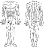
Emergency War Surgery NATO Handbook: Part IV: Regional Wounds and Injuries: Chapter XXXIII: Wounds and Injuries of the Spinal Column and Cord Anatomical ConsiderationsUnited States Department of Defense Injury to the upper cervical spinal cord between C-1 and C-4, the level from which the cervical plexus and the phrenic nerves are derived, can result in the loss of both voluntary and involuntary diaphragmatic motion, the loss of chest wall muscle function, and the loss of function of the cervical strap muscles, which serve as accessory muscles of ventilation. A complete injury at this level, in the absence of some method of immediate assist, results in cessation of ventilation and death. When the cervical cord is injured below this level, the level of the cord injury is determined by assessment of motor and sensory function (Figure 40 and Table 16). The presence of any neural function below the level of the bony injury, to include the-preservation of motor or sensory activity within the perianal (S-2, S-3) sacral dermatomes (sacral sparing), indicates an incomplete cord injury and is a favorable prognostic sign. Axial loading (compression) injuries of the upper cervical spine can cause disruption of the ring of C-1 (Jefferson fracture). This fracture is rarely accompanied by cord damage because of the width of the neural canal at the C-1 and C-2 levels.* A C-1 fracture is usually stable and can be managed nonoperatively in the absence of other fractures or signs of instability. An associated fracture of C-2 must always be ruled out when fractures of C-1 are present. In this case, management depends on the type of injury present at C-2. Odontoid process (dens) fractures involving C-2 occur along the process in one of three locations. Type I fractures pass through the uppermost portion of the dens. Type II fractures of the odontoid pass through the base of the dens. Since the upper and lower segments are attached to opposing ligamentous and bony structures, there usually is separation and these fractures are unstable. Type III fractures occur at the junction of the dens and body of C-2. Type I and Type III fractures are normally stable and can be managed with immobilization only. Type II fractures are unstable and require surgical stabilization. These fractures must be stabilized during the assessment phase with Gardner Wells skeletal traction followed, in time, by either halo or other orthopedic apparatus, fixation or surgical stabilization with early internal wire, or plate and screw fixation. Axial load forces applied to the head and upper cervical spine may disrupt the posterior elements of C-2 (Hangman's fracture). This is a relatively stable fracture and is usually managed nonoperatively. When fracture of the posterior elements of C-2 is accompanied by displacement, dislocation, or fracture of the body of C-2, surgical stabilization is indicated. Fractures or dislocations of the cervical spine between C-3 and C-7 are caused by hyperflexion, axial load, rotation, or a combination of these forces. Typically these injuries result in instability. Hyperextension injuries to the cervical spine usually occur at the C-6, C-7 interspace, but produce complete neurological injuries less often than do flexion injuries. The extent of the injury depends on how much ligamentous and vertebral element integrity (two column integrity) is lost. The severity of the skeletal injury and the resulting neurological deficit do not always correlate. Facet joint fractures and dislocations are associated with flexionrotation injuries. They are often difficult to demonstrate on the initial anterior-posterior and lateral radiographs. For this reason, tomographic studies may be necessary. Thirty percent displacement of one vertebra on another indicates unilateral facet dislocation, whereas 50% displacement indicates bilateral facet disruption. Unilateral facet disruption is usually stable. In the absence of neurological findings, this injury can be managed nonoperatively. If it does not reduce with traction, this injury should be surgically reduced and stabilized. Bilateral facet dislocations are always unstable and require surgical stabilization. Complete neurological injury normally accompanies this injury. *One-Third Rule: At this level one-third of the spinal canal is occupied by the spinal cord, one-third by the odontoid process, and one-third is free space. The vascular supply of the spinal cord is most vulnerable between T-4 and T-6, where the neural canal is most narrow. Even minor degrees of vertebral column malalignment in this region result in neurological injury. Thoracic cord injury usually results from a combination of flexion, axial loading, and rotation forces. These stress forces are seen with parachute jumps and pilot ejections from highperformance aircraft. While the thoracic rib cage contributes to the rotary stability of the thoracic spine, wedge compression (flexion) fractures of the upper thoracic vertebral column are not uncommon. The most common site for a compression fracture is at L-1 and L-2. When not accompanied by other elements of injury, anterior wedge compression fractures of 25-30 % can be considered stable Greater degrees of compression and associated displacement require surgical stabilization. Most axial-loading burst fractures in the lumbar region occur between L-2 and L-4 and are unstable. These fractures often cause extrusion of bone into the spinal canal and/or progressive angular deformity. Surgical stabilization and, occasionally, removal of bone fragments that compress the spinal cord constitute the definitive management of these injuries.
Approved for public release; Distribution is unlimited. The listing of any non-Federal product in this CD is not an endorsement of the product itself, but simply an acknowledgement of the source. Operational Medicine 2001 Health Care in Military Settings
This web version is provided by The Brookside Associates Medical Education Division. It contains original contents from the official US Navy NAVMED P-5139, but has been reformatted for web access and includes advertising and links that were not present in the original version. This web version has not been approved by the Department of the Navy or the Department of Defense. The presence of any advertising on these pages does not constitute an endorsement of that product or service by either the US Department of Defense or the Brookside Associates. The Brookside Associates is a private organization, not affiliated with the United States Department of Defense. |
