|
|
Medical Education Division |
Operational Medicine 2001
Emergency War Surgery
Second United States Revision of The Emergency War Surgery NATO
Handbook
United States Department of Defense
Emergency War Surgery
Second United States Revision of The Emergency War Surgery NATO Handbook
United States Department of Defense
Home À Military Medicine À Sick Call À Basic Exams À Medical Procedures À Lab and X-ray À The Pharmacy À The Library À Equipment À Patient Transport À Medical Force Protection À Operational Safety À Operational Settings À Special Operations À Humanitarian Missions À Instructions/Orders À Other Agencies À Video Gallery À Phone Consultation À Forms À Web Links À Acknowledgements À Help À Feedback
|
Emergency War Surgery NATO Handbook: Part IV: Regional Wounds and Injuries: Chapter XXIV: Wounds and Injuries of the Eye Management: Major Injuries: Forward HospitalUnited States Department of Defense Forward Hospital - In the absence of an ophthalmologist, treatment of major ocular injuries in forward hospitals normally is managed by the general surgeon and ideally is limited to interim measures aimed at prevention of infection within the eye. Systemic antibiotics and tetanus prophylaxis should be instituted at the earliest opportunity in the preoperative period. Lid and conjunctival debris should be carefully irrigated away. Any sterile irrigating solution, including water, is acceptable. This should be followed by generous topical application of fresh solutions of an ophthalmic antibiotic (gentamycin sulfate, chloramphenicol or neomycin sulfatepolymixin B sulfate) and atropine sulfate 1%. A sterile, four-by-four-inch gauze strip is applied to keep the area clean, and additional protection is afforded by taping a Fox (or similar type) shield over the injured eye. A pressure dressing should be avoided as it may cause serious damage by expressing intraocular contents through a penetrating wound. Since patching also helps provide an excellent culture medium for bacteria, particularly Pseudomonas, topical antibiotic solution is carefully reinstilled every four hours, and a fresh, sterile gauze patch reapplied twice daily. Sterile irrigation of mucopurulent secretions from the lid margins and conjunctiva should be carried out when the gauze dressing is changed. The uninjured eye should be patched to reduce unwanted ocular motion. No ocular surgery should be performed. Particularly, no attempt should be made to remove protruding or penetrating foreign bodies or to repair corneal or scleral lacerations. Preferably, repair should be undertaken for lacerations involving the lid margin or the nasolacrimal apparatus. Even an eye which appears grossly irreparable may have surgery deferred, utilizing the same regimen of sterile gauze dressings and antibiotics. Until recently, the selection of systemic antibiotics has been beset with two problems: (1) many drugs do not pass the blood-aqueous and blood-retina barriers to give adequate intraocular tissue concentrations, and (2) earlier drugs have had limited bactericidal spectra, especially for strains of Pseudomonas aeruginosa. When ophthalmologic care must be delayed, the following initial antibiotic regimen may be used if infection is suspected and the wound is of such size and location that extrusion of intraocular contents is not a risk: Subconjunctival: Gentamycin 40mg
Cephaloridine 100 mg
or
Gentamycin 40mg
Methicillin 100 mg
Topical: Gentamycin 9mg/cc
Bacitracin 5,000 u/cc
Systemic: Cephaloridine, 1 gm. stat, IV then 500mg q 6 hr.
or
Methicillin 2gm, IM, q 8 hr.
Subconjunctival injection is best accomplished using topical proparacaine (0.5%) anesthesia, a smallvolume syringe (2.5cc) and a short (5/8") 27 gauge needle. The bulbar conjunctiva is engaged near the upper or lower fornix with the bevel facing the globe, and the needle is advanced toward the fornix, the injection being given while the needle tip is visible through the conjunctiva. Subconjunctival injections are contraindicated if the wound is of such size and location as to risk extrusion of intraocular contents. In such cases, only the topical and systemic routes should be used, as noted above. While ideally handled by an ophthalmologist, many of the following ocular injuries can be managed well by surgeons or general medical officers:
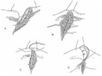
If evacuation or ophthalmologic care is delayed, repair of lid lacerations by a non-ophthalmologic surgeon may be necessary. Evaluation of any lid injury must include an evaluation for coexisting injury to the eyeball and penetrating injury to the intracranial contents. Lacerations and avulsions near the medial canthal tendon necessitate a careful examination for interruption of the canaliculus. In the repair of any lid injury, it is necessary respect the complex anatomy of the lid, exact anatomical realignment being necessary (Figure 31). It is especially important that the levator muscle, the tarsal plate, and the medial canthal tendon be precisely reapproximated, or severe functional and cosmetic disabilities may ensue. Adequate coverage of the cornea is of critical importance. The repair of lid injuries requires a knowledge of the anatomy of the lid, fine ophthalmic instruments and sutures, and magnification provided by either loupes or an operating microscope. Lid tissue should be preserved wherever possible. Only tissue that is clearly necrotic should be debrided. Totally avulsed lid segments should be reattached after cleansing. Lacerated lids should be extensively irrigated and all foreign bodies removed. Lid lacerations should be repaired in the following manner. Lacerations through the skin horizontal to the lid margin can be repaired with 6-0 black silk sutures. Lacerations that involve the lid margin itself must be repaired precisely: 4-0 black silk suture should be used to approximate the tarsal plates elsewhere and 6-0 black silk should be used to approximate the anterior and posterior borders of the lid margin and the skin of the lid elsewhere. Lid margin sutures should stay in for ten days. The lid should be placed on stretch using the long arms of the 4-0 black silk sutures for at least three days after the repair of the injury. A light pressure dressing should be placed over the eye after the instillation of an antibiotic ointment. The cornea must be checked each day. No elaborate reconstruction of the lids should be performed in a combat zone, though every effort should be made to preserve and reapproximate lid tissues at the time of the primary repair.
Approved for public release; Distribution is unlimited. The listing of any non-Federal product in this CD is not an endorsement of the product itself, but simply an acknowledgement of the source. Operational Medicine 2001 Health Care in Military Settings
This web version is provided by The Brookside Associates Medical Education Division. It contains original contents from the official US Navy NAVMED P-5139, but has been reformatted for web access and includes advertising and links that were not present in the original version. This web version has not been approved by the Department of the Navy or the Department of Defense. The presence of any advertising on these pages does not constitute an endorsement of that product or service by either the US Department of Defense or the Brookside Associates. The Brookside Associates is a private organization, not affiliated with the United States Department of Defense. |