|
Emergency War Surgery NATO Handbook: Part III: General Considerations of Wound
Management: Chapter XVI: Wounds and Injuries of the Soft Tissues
Treatment Recommendations
United States Department of Defense
Establish an adequate blood level of penicillin or an antibiotic with a similar
spectrum as soon as possible after wounding. Make generous incisions of the wound to
relieve mechanical pressure and establish open drainage. Remove easily-accessible foreign
bodies and detached pieces of muscle, and irrigate the wound copiously. The wound track is
then inspected and any additional muscle whose gross architecture is severely disrupted is
excised. At the conclusion of the procedure, complete hemostasis must be achieved to
preclude the subsequent development of collections within the wound that would impede
capillary perfusion of borderline tissues. The technique is shown in Figure 24.
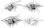
Figure 24
-
Excise entrance and exit wounds with a narrow margin of skin oriented parallel to the
underlying muscle fibers. This excised skin margin should include, in continuity, the
underlying subcutaneous tissue. These incisions should be generous, such that optimal
surgical exposure and adequate subsequent drainage will be achieved.
-
Through these openings, generously incise the fascia parallel to the muscle fibers in
both directions. The underlying muscle surrounding the missile tract should be opened in
the direction of its fibers to the degree necessary to achieve exposure adequate to
inspect the track, remove foreign bodies, and excise non-viable muscle These maneuvers are
performed at both the wound of entry and the exit wound. The muscle surrounding the
central portion of the track can usually be dealt with through the entry and exit wounds.
For example, a mid-thigh, through-and-through wound of the soft tissues can generally be
surgically managed by working through the excised and extended wounds of entry and exit.
This approach precludes the necessity of cutting across good muscle groups as is generally
the case when one elects to connect the two wounds. Appropriate drainage of war wounds is
often easier said than done. Liberal incisions tend to facilitate drainage from the
wound's deeper recesses. Whereas excision of skin, fascia, arteries, nerves, veins, and
bone is conservative, the excision of muscle should be more liberal.
-
As a dressing, dry sterile gauze should be laid lightly in the wound. This should be no
more than a wick. In no case should gauze be "packed" into the wound since this
additional pressure can cause necrosis of any tissue that already has its blood supply
partially compromised.
-
The single most important principle in the management of battle wounds is their
nonclosure following debridement. The surgeon must not give in to the temptation to
primarily close certain "very clean appearing" war wounds. Such closure is ill
advised and inappropriate and can only be condemned. All wounds must be left widely open
with the following exceptions:
-
Sucking chest wounds
-
Joint capsules
-
Wounds of the dura
-
Some head and neck wounds; however, with severe contamination it may be safer to leave
these open.
-
The delayed primary wound closure is usually performed in a communication zone hospital
4-10 days after debridement, but occasionally may be performed at the forward hospital
when evacuation has had to be delayed. The indication for delayed primary closure is the
clinically clean appearance of the wound. Whereas most wounds are closed in the operating
room utilizing the interrupted wire technique and local or general anesthesia, some may be
very amenable to tape closure. This technique can be initiated 4-6 days post debridement.
Approximation of the skin edges is accomplished with micropore paper tape or wide
"butterflies" applied in overlapping diagonal "basket weave" fashion
after the skin has been degreased with acetone, and tincture of benzoin has been applied
and allowed to dry thoroughly. Edges of the wound may not come completely together with
the first tape application. This is not a problem, as they will come progressively closer
together with each reapplication of tape, done at 48 hour intervals. Tape closure offers
some advantages over suture closure Even compression of wound edges decreases skin edema,
and the problem of cutting needles causing additional tissue damage is avoided. The wound
edges are very vascular and needle passage can cause hematomas. Since tape closure is, in
reality, a gradual "encouragement" of the skin toward closure rather than a
total closure from the beginning, a great margin for error is added and the potential
complication of wound breakdown, sometimes seen after suture closure, is almost completely
avoided. No anesthesia is needed for this procedure and it can be performed by supervised
ward nursing personnel.
It should be recognized that even though the surgeon diligently
attempts to excise all devitalized tissue, the dynamics of wound physiology and the
imperfections of ones ability to absolutely identify nonviable tissue are such that some
devitalized muscle may be left behind or evolve over time in the wound. In the
appropriately drained wound, this minimal amount of devitalized tissue will be absorbed or
extruded. A small percentage of these wounds will require a second debridement prior to
delayed primary closure. At worst, in the absence of adequate drainage, an abscess that
requires subsequent drainage may develop. In this situation, antibiotics localize or
isolate the deleterious effects of the injury to the site of injury, thereby precluding
systemic, lifethreatening sepsis.
Approved for public release; Distribution is unlimited.
The listing of any non-Federal product in this CD is not an
endorsement of the product itself, but simply an acknowledgement of the source.
Operational Medicine 2001
Health Care in Military Settings
Bureau of Medicine and Surgery
Department of the Navy
2300 E Street NW
Washington, D.C
20372-5300 |
Operational Medicine
Health Care in Military Settings
CAPT Michael John Hughey, MC, USNR
NAVMED P-5139
January 1, 2001 |
United States Special Operations Command
7701 Tampa Point Blvd.
MacDill AFB, Florida
33621-5323 |
This web version is provided by
The Brookside Associates Medical
Education Division. It contains original contents from the official US
Navy NAVMED P-5139, but has been reformatted for web access and includes
advertising and links that were not present in the original version. This web
version has not been approved by the Department of the Navy or the Department of
Defense. The presence of any advertising on these pages does not constitute an
endorsement of that product or service by either the US Department of Defense or
the Brookside Associates. The Brookside Associates is a private organization,
not affiliated with the United States Department of Defense.
Contact Us À À
Other Brookside Products
|