|
Medical Education Division |
Operational Medicine 2001
Field Medical Service School
Student Handbook

EMERGENCY CRICOTHYROIDOTOMY
FMST
0410 17
Dec 99
Terminal
Learning Objectives: 1.
Given an
upper airway obstruction in a combat environment (day and night) and the
standard Field Medical Service Technician supplies and equipment, perform
emergency cricothyroidotomy, per the references. (FMST .04.11) Enabling
Learning Objectives: 1.
Without
the aid of reference materials and given a diagram, identify important
anatomical landmarks of the neck and throat, per the student handbook.
(FMST .04.11a) 2.
Without
the aid of reference materials, identify the purposes and indications for
performing an emergency cricothyroidotomy, per the student handbook.
(FMST .04.11b) 3.
Without
the aid of reference materials and given a list, select the complications which
may result from performing an emergency cricothyroidotomy, per the student
handbook. (FMST .04.11c) 4.
Without
the aid of reference materials and given a FMST MOLLE Medic bagand simulated
casualty, perform an emergency cricothyroidotomy, per the student handbook.
(FMST .04.11d) OUTLINE:
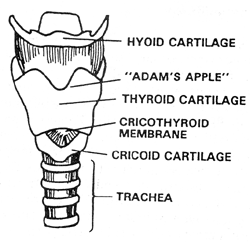
A. ANATOMY 1.
Trachea – also known as the windpipe.
It is the cartilagenous and membranous tube descending from, and
continuous with, the lower part of the larynx to the bronchi. 2.
Thyroid Cartilage – also known as the “Adam’s Apple.”
The thyroid cartilage is located in the upper part of the throat.
The thyroid cartilage tends to be more prominent in men than women. 3.
Cricoid Cartilage – located approximately three quarters of an inch
(3/4”) inferior to the thyroid cartilage.
It is a firm ridge which is one-eighth to one quarter of an inch thick.
The cricoid and thyroid cartilage form the framework of the larynx. 4.
Cricothyroid Membrane – the soft tissue depression between the thyroid
and cricoid cartilage. This
membrane connects the two cartilages and is only covered by skin.
5.
Carotid Arteries – the two principal arteries of the neck. 6.
Jugular Veins – the two principal veins of the neck. 7.
Esophagus – The musculomembranous tube extending down downward from the
pharynx to the stomach. The
esophagus lies posterior to the trachea. 8. Thyroid Gland - The largest exocrine gland in man, the thyroid gland is situated in front of the lower part of the neck. It consists of a right and left lobe on either side of the trachea.
B. EMERGENCY CRICOTHYROIDOTOMY - THEORY
a.
Obstructed airway – an obstructed object will usually prevent the
passage of an endotracheal tube through the airway. Therefore, a surgical airway
distal to the obstruction is required. Causes
of an obstructed airway include: 1.
Facial and oropharyngeal edema from inhalation burns 2.
Facial and oropharyngeal edema from anaphylaxis 3.
Foreign objects b.
Congenital deformities of the oropharynx or nasopharynx which inhibit or
prevent nasotracheal or orotracheal intubation. c.
Trauma to the head and neck which would preclude the use of an ambu-bag,
oropharyngeal airway, nasopharyngeal airway, and endotracheal tube insertion.
Examples include: 1.
Facial and oropharyngeal edema from severe trauma 2.
Facial fractures (mandible fracture) d.
Cervical spine fractures in a patient who needs an airway but whom
nasotracheal intubation is unsuccessful or contraindicated.
Examples include: 1.
Nasal bone fractures 2.
Cribiform fractures e.
The healthcare provider is unable to establish an airway by any other
means and this is the “last resort.”
a.
Massive trauma to the larynx or cricoid cartilage – damage to the
affected structures will make it impossible to perform the procedure properly b.
Contraindicated if another means of establishing an airway have not been
attempted (i.e. nasotracheal or orotracheal intubation)
a.
Provides a definitive airway for ventilating the patient b.
Can be performed quickly and has few complications associated with the
procedure
a.
Need advanced training to properly perform procedure b.
Bypasses the nares function of warming and filtering the air c.
May increase respiratory resistance d.
Improper placement
a.
HEMORRHAGE – is the most common complication 1.
Definition – the loss of a large amount of blood in a short amount of
time 2.
Causes a)
Minor bleeding – caused by lacerating superficial capillaries in the
skin tissue b)
Significant bleeding – caused by lacerating major vessels (carotid
artery and the jugular veins) within the neck 3.
Treatment a)
Minor - direct pressure to control the bleeding then apply a simple
pressure dressing b)
Significant – same as minor. However,
if unable to control the bleeding, the vessel may need to be ligated. b.
ESOPHAGEAL PERFORATION OR TRACHEOESOPHAGEAL FISTULA 1.
Definition – the creation of a
hole between the esophagus and trachea 2.
Causes a)
Creating an incision too deep through the cricothyroid membrane b)
Forcing the endotracheal tube through the cricothyroid membrane and into
the esophagus 3.
Treatment – requires surgical repair at higher echelon of care c.
SUBCUTANEOUS EMPHYSEMA 1.
Definition – the presence of free air or gas within the subcutaneous
tissues 2.
Causes a)
Creating too wide of an incision will encourage air entrapment under the
subcutaneous tissue b)
Air leaking out of the insertion site may get trapped under the
subcutaneous tissues 3.
Treatment a)
No treatment is necessary. The
subcutaneous emphysema will dissipate on its own accord within a few days. b)
Placing a petroleum gauze dressing around the incision / insertion site
will help reduce the incidence of subcutaneous emphysema. c)
Monitor size of subcutaneous emphysema C.
EMERGENCY CRICOTHYROIDOTOMY – PROCEDURAL STEPS
a.
#11 Scalpel Blade b.
Scalpel Blade Handle c.
Endotracheal tube – shortened d.
10 cc syringe – used to fill the cuff at the end of the endotracheal
tube e.
Stylet – a wire inserted into the endotracheal tube in order to stiffen
the tube during passage f.
Water Soluble Lubrication – KY Jelly or Surgilube g.
Stethoscope – to check for proper placement of the endotracheal tube h.
Curved Kelly Hemostat – used to open the incision site i.
Tissue Forceps – used to retract skin tissue at the incision site j.
Ambu-bag – to ventilate patient k.
Sterile Dressing l.
Petroleum Gauze m.
Betadine or Alcohol Wipes n.
Sterile or Clean Gloves o.
Suture material p.
Suction device q.
Suture Scissors r.
Tape
a.
Maintain the patient’s ABC’s b.
Determine that the patient requires an emergency cricothyroidotomy c.
Assemble required equipment d.
Position the patients head 1.
The patient is placed in a supine or semi-recumbent position 2.
The neck is placed in a neutral position e.
Palpate the thyroid and cricoid cartilage for orientation f.
Locate the cricothyroid membrane g.
Stabilize the thyroid cartilage using your non-dominant hand h.
Swab the incision site with alcohol or betadine swabs i.
Make a vertical incision through the skin approximately 2-5 cm (1 inch)
long over the cricothyroid membrane j.
Visualize the cricothyroid membrane k.
Make a transverse incision into the cricothyroid membrane 1.
Do not make the incision more than ½ inch deep or you may perforate the
esophagus l.
Insert Curved Kelly Hemostat into incision and blunt dissect incision
(turn the Curved Kelly Hemostat 90degrees to open up the incision) m.
Insert the shortened endotracheal tube into the incision, directing the
tube distally down the trachea n.
Ventilate the patient with two breaths 1.
Check for proper placement with these first two ventilation’s by: a)
Observing the chest rise and fall with each ventilation b)
Auscultate for bilateral breath sounds 1)
Bilateral Breath Sounds Present – the endotracheal tube has been
properly placed: (a)
Proper placement will cause both lungs to inflate with each ventilation 2)
Bilaterally Absent Breath Sounds -
the endotracheal tube is not within the trachea and has probably been placed
within the esophagus. (a)
Remove the endotracheal tube and attempt to reinsert into the trachea 3)
Breath Sounds in Right Lung Field – the endotracheal tube has been
placed too far down the bronchial tree and is in the right mainstem bronchus.
(a) Pull
back the endotracheal tube ¼ - ½ inch or until bilateral breath sounds have
been established c)
Auscultate over epigastrium for gastric sounds 1)
Placement of the endotracheal tube into the stomach / esophagus will produce
gurgling sounds in the epigastric area with ventilations o.
Inflate the endotracheal tube’s cuff with 10 cc’s of air 1.
Inflation of the balloon cuff serves two purposes: a)
Holds the endotracheal tube in place b)
Acts as a barrier and prevents fluids from entering the lungs p.
Ventilate the patient with two breaths of 100% oxygen q.
Suture the endotracheal tube in place (if required) r.
Apply petroleum gauze dressing to insertion site s.
Apply dry sterile dressing to insertion site
t.
Continue to ventilate patient (1 breath every 5 seconds) and suction as
necessary u.
Continue to monitor the patient for changes
REFERENCE
(S): 1.
EMERGENCY WAR SURGERY 2.
TEXTBOOK OF ADVANCED CARDIAC LIFE SUPPORT 3.
PREHOSPITAL EMERGENCY CARE AND CRISIS INTERVENTION
Field Medical Service School
Approved for public release; Distribution is unlimited. The listing of any non-Federal product in this CD is not an endorsement of the product itself, but simply an acknowledgement of the source. Operational Medicine 2001 Home · Military Medicine · Sick Call · Basic Exams · Medical Procedures · Lab and X-ray · The Pharmacy · The Library · Equipment · Patient Transport · Medical Force Protection · Operational Safety · Operational Settings · Special Operations · Humanitarian Missions · Instructions/Orders · Other Agencies · Video Gallery · Phone Consultation · Forms · Web Links · Acknowledgements · Help · Feedback
*This web version is provided by The Brookside Associates Medical Education Division. It contains original contents from the official US Navy NAVMED P-5139, but has been reformatted for web access and includes advertising and links that were not present in the original version. This web version has not been approved by the Department of the Navy or the Department of Defense. The presence of any advertising on these pages does not constitute an endorsement of that product or service by either the US Department of Defense or the Brookside Associates. The Brookside Associates is a private organization, not affiliated with the United States Department of Defense. |


