This is the Archived Desktop Edition.
You should be transferred to the Newest Edition for Desktop and Mobile within 2 seconds.
![]()
Multimedia Edition
|
Lesson 6: Complications of Postpartum
Section III. COMPLICATIONS OF POSTPARTUM
Postpartal hemorrhage is the postpartum loss of blood totaling 500 ml or more within a twenty-four hour period. After bladder distention is ruled out, the three main causes of postpartal hemorrhage are uterine atony, lacerations, and retained placental fragments in the uterus.
a. Uterine Atony. This is the inability of the myometrium to contract and constrict the blood vessels within the muscle fibers, resulting in open sinuses at the site of placental separation. Decreased muscle tone causes slow, insidious loss of blood.
(1) Factors usually leading to uterine atony.
(a) Conditions which result on overextension of uterine musculature (multiple pregnancy - two or more fetuses and hydramnios - excessive amniotic fluid).
(b) Conditions resulting in exhaustion of the uterine musculature are large fetuses, prolonged or difficult labor, Pitocin® induced or augmented labor (this may result in decreased response to postpartal administration of pitocin) and precipitous or forceful delivery.
(2) Situations resulting in drug related relaxation of uterine musculature are the use of MgSO4 for preeclampsia and the use of general anesthesia for cesarean delivery. Conditions resulting in abnormal bleeding or uterine tissue damage are cesarean section, placenta previa, abruptio placenta, uterine rupture, and retained placental fragments.
(3) Signs and symptoms of uterine atony.
(a) Signs of shock--decreased blood pressure, increased pulse, and increased and anxiety and irritability.
(b) Bleeding-usually dark with clots present.
(c) Noncontracted, boggy uterine fundus.
(4) Medical treatment.
(a) Intervenously fluids administered to increase fluid and blood volume.
(b) Oxytocin administration.
(c) Methergine/prostin may be administered to stimulate uterine contractions when oxytocin is ineffective.
(d) Blood transfusion if the patient's hematocrit drops too low and/or if she is symptomatic.
(5) Nursing interventions.
(a) Palpate the fundus frequently to determine continued muscle tone.
(b) Massage the fundus, if boggy, until firm (do not over massage, this fatigues the muscle).
(c) Monitor patient's vital signs every 15 minutes until stable.
(d) Prevent bladder distention. Bladder distention displaces the uterus and prevents effective uterine contractions.
(1) Common sites. Sites of lacerations are the vaginal side wall, the cervix, the lower uterine segment, and the perineum.
(2) Degrees of perineal lacerations.
(a) First degree-tear of the vaginal and perineal mucous membranes.
(b) Second degree-tear of the vaginal and perineal mucous membrane and the perineal muscles.
(c) Third degree-tear of the vaginal and perineal mucous membrane, the perineal muscles, and the capsule of the rectal sphincter.
(d) Fourth degree-tear of the vaginal and perineal mucous membrane, the perineal muscles, and through the rectal sphincter and anterior wall of the rectum.
(3) Possible causes.
(a) Rapid descent of the fetus.
(b) Pushing prior to complete cervical effacement and dilatation.
(c) Large fetus.
(d) Forceps application.
(e) Uncontrolled, forceful extension of the fetal head.
(4) Signs and symptoms.
(a) Obvious body injury after delivery of the infant--if perineal laceration.
(b) Bright red bleeding despite a well toned fundus-if vaginal or cervical laceration and not detected at time of delivery.
(c) Signs of shock-rapid, thready pulse, falling blood pressure, increasing anxiety of the patient.
(5) Medical treatment.
(a) Suturing of the laceration.
(b) Vaginal packing.
(c) Blood transfusions if the patient's hematocrit is low and the patient is symptomatic.
(6) Nursing interventions.
(a) Observe closely for continued vaginal bleeding.
(b) Monitor the patient's vital signs.
(c) Flag the patient's chart for vaginal packing in place. This is helpful to the nurse who is checking for vaginal bleeding doesn't mistake a lack of obvious signs of blood for no bleeding. The vaginal packing could "hide" a hemorrhagic episode of bleeding.
c. Retained Placental Fragments in the Uterus. These fragments are the major cause of late postpartum hemorrhage.
(1) Signs and symptoms.
(a) Large amount of bright red bleeding or persistent trickle type bleeding.
(b) Uterus may be boggy due to its inability to contract properly.
(c) Signs of shock.
(d) Sudden rise in uterine fundal height indicating the formation of clots inside the uterine cavity.
(2) Medical treatment.
(a) Manual removal of the remaining placenta is done by the physician, if it is a result of incomplete separations of the placenta with increased vaginal bleeding.
(b) A D&C is performed, if it is retained fragments.
(c) Intravenous fluids are administered.
(d) Oxytocic drugs are given immediately after either procedure.
(3) Nursing interventions.
(a) Check the uterine fundus tone frequently (every 15 minutes the first hour, then every 30 minutes for 2 hours, and every hour until stable).
(b) Check the nature and amount of lochia flow (every 15 minutes the first hour, then every 30 minutes for 2 hours, and every hour until stable).
(c) Keep accurate count of perineal pads used.
(d) Monitor the patient's vital signs and blood pressure every 15 minutes or more frequently as necessary.
(e) Observe for signs of shock.
(f) Turn the patient on her side to prevent pooling of blood under her.
(g) Provide emotional support to the patient and family.
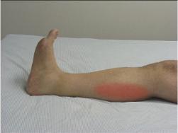
Vulvar hematoma is a localized collection of blood in the connective tissue beneath the skin covering the external genitalia or vaginal mucosa. It generally forms as a result of injury to the perineal blood vessels during the delivery process.
a. Causes of Hematomas.
(1) Rapid, spontaneous delivery.
(2) Perineal varicosities.
(3) Episiotomy repairs.
(4) Laceration of perineal tissues.
b. Signs and Symptoms.
(1) Severe, sharp perineal pain.
(2) Appearance of a tense, sensitive mass of varying size covered by discolored skin.
(3) Swelling in the perineal wall.
(4) Often seen on the opposite side of the episiotomy.
(5) Inability to void due to pressure/edema on or around the urethra.
(6) Complaint of fullness or pressure in the vagina.
c. Medical Treatment. This is consists of analgesics given for discomfort, opening the hematoma so blood clots can be evacuated and the bleeders can be ligated, and packing for pressure.
d. Nursing Interventions.
(1) Apply ice to area of hematoma.
(2) Observe for evidence of enlarged hematoma.
(3) Flag the patient's chart if packing was inserted.
Uterine subinvolution is a slowing of the process of involution or shrinking of the uterus.
a. Causes. Endometritis, retained placental fragments, pelvic infection, and uterine fibroids may cause uterine subinvolution.
b. Signs and Symptoms.
(1) Prolonged lochial flow.
(2) Profuse vaginal bleeding.
(3) Large, flabby uterus.
c. Medical Treatment.
(1) Administration of oxytocic medication to improve uterine muscle tone. Oxytocic medication includes
(a) Methergine®-a drug of choice since it can be given by mouth.
(b) Pitocin®.
(c) Ergotrate®.
(2) Dilation and curettage (D&C) to remove any placental fragments.
(3) Antimicrobial therapy for endometritis.
d. Nursing Interventions.
(1) Early ambulation postpartum.
(2) Daily evaluation of fundal height to document involution.
Puerperal infection is a term used to describe any infection of the reproductive tract during the first six weeks of postpartum.
|
a. Pathology. When the third stage of labor is completed, the placental attachment site is raw, elevated, and dark red. The surface is nodular, owing to the numerous veins, and offers an excellent portal of entry for microorganisms. The uterine decidua is very thin and has many small openings that offer a portal for pathogens. In addition, small cervical, vaginal and perineal lacerations, as well as the episiotomy site, provide entry ports for pathogens. The resultant inflammation and infection can remain localized or can extend via blood or lymph vessels to other tissues.
b. Organisms. Those organisms recognized as the common causative agents are normally seen in the lower bowel and lower genital tract.
(1) Anaerobic staphylococci.
(2) Anaerobic streptococci.
(3) Clostridium perfringens.
(4) Neisseria gonorrhea.
c. Predisposing Factors.
(1) Prolonged rupture of uterine membranes provides increased opportunity for infection to develop prior to delivery.
(2) Retained placental fragments-provides additional medium for infectious growth.
(3) Postpartal hemorrhage-causes decreased resistance to pathogens.
(4) Preexisting anemia-low resistance to infection.
(5) A prolonged and difficult labor, especially with the involvement of instruments (forceps).
(6) Intrauterine manipulations for fetal delivery or manual expulsion of placenta.
d. Spread of Infectious Microorganisms. This may be the result of the spread of infectious microorganisms in the hospital setting.
e. Means to Prevent the Spread of Puerperal Infection in Hospitals.
(1) Restrict personnel with respiratory infections from working with patients.
(2) Use caps, mask, gowns, and gloves when working in delivery rooms.
(3) Use sterilized equipment within control dates.
(4) Wash hands meticulously (staff).
(5) Correct breaks in sterile techniques immediately.
(6) Instruct the patient on hand washing and cleansing her perineum from front to back.
(7) Limit unnecessary vaginal exams during labor which increases the chances of introducing organisms from the rectum and vagina into the uterus.
f. Kinds of Postpartal Infections.
(1) Endometritis-invasion of microorganisms into the placental site of the uterine wall.
(2) Pelvic cellulitis (parametritis)-infection that has spread beyond the endometrium into the surrounding pelvic structures including the broad ligament.
(3) Peritonitis-an infection of the peritoneum, either generalized or localized.
(4) Salpingitis-an infection of the fallopian tubes following childbirth.
g. Medical Treatment of Puerperal Infection.
(1) Antibiotics to which the causative organisms are sensitive, analgesics, and sedatives.
(a) Initial antibiotics are given by IV until the fever resolves.
(b) May possibly switch from IV and give oral medication if fever remains normal for 48 to 72 hours.
(c) May use a course of triple antibiotics until all cultures are obtained.
(2) Incision and drainage (I&D) of any abscesses formed.
h. Nursing Care of Puerperal Infection.
(1) Isolation, if possible, the removal of the patient from the maternity ward.
(2) Meticulous hand washing.
(3) Patient placed in Fowler's position to facilitate drainage.
(4) Reeducation of the patient on handwashing and peri-care.
(5) Emotional support since the patient may be prevented from rooming in with her infant while her temperature is elevated.
a. General. Thrombophlebitis is an inflammation/infection of pooled and clotted blood in a vein.
(1) Types of Thrombophlebitis.
(a) Femoral- inflammation along the femoral, popliteal, or saphenous veins.
(b) Pelvic-inflammation/infection of the pelvic veins.
(c) Superficial- inflammation/infection of the superficial saphenous veins.
(2) Signs and Symptoms.
(a) Pain.
(b) Fever.
(c) Localized tenderness and/or swelling and redness.
(d) Chills.
(3) Medical Treatment.
(a) Antibiotic therapy.
(b) Anticoagulant therapy-heparin.
(c) Blood transfusions as needed.
(4) Nursing Management.
(a) Bed rest.
(b) Analgesics as needed.
(c) Elastic leg supports where indicated.
(d) For leg involvement, apply warm moist soaks to affected area(s).
|
b. Pulmonary Embolus. This is a major complication of thrombophlebitis. It results when a clot breaks loose, travels through the circulatory system, and obstructs the pulmonary arterial bed. It is a serious, life-threatening situation.
(1) Signs and symptoms.
(a) Chest pain.
(b) Sudden shortness of breath.
(c) Rapid respirations.
(d) Air hunger/anxiety.
(e) Circulatory collapse--weak, rapid pulse and hypotension.
(f) Cyanosis.
(2) Treatment and nursing care.
(a) Administer oxygen as ordered.
(b) Give sedatives to relax the patient as ordered.
(c) Perform surgery to remove the embolus.
(d) Monitor vital signs very closely (at least every hour).
(e) Transfer to intensive care unit (ICU) if necessary.
(f) Provide emotional support since the patient may be restricted from seeing her baby due to visitation policies.
Mastitis is inflammation of the breast tissue, usually unilateral after the milk flow is established. It is caused by streptococcal or staphylococcal invasion of the breast tissue through cracks or fissures around the nipple. It may be obtained from the infant's nose or throat. The infant probably acquired it while in the nursery.
a. Signs and Symptoms.
(1) Erythema over the infected breast.
(2) Marked breast engorgement.
(3) Acute breast pain, tenderness.
(4) Fever and chills.
(5) Ancillary lymph gland enlargement.
b. Medical Treatment.
(1) Antibiotic therapy and analgesic therapy.
(2) Periodic cultures of breast milk.
(3) Intravenous fluids.
(4) Possible I&D, if abscesses.
(5) Discontinued breast-feeding for a short time depending on antibiotic used and closeness of abscess site to nipple.
c. Nursing Care.
(1) Apply ice or heat to painful, swollen breast depending on the stage of infection. Ice should be avoided if the mother plans to resume or continue breast-feeding.
(2) Encourage increased fluids.
(3) Inform mother to wear a support bra.
(4) Have the mother pump her breast until nursing resumes. Pumping the breast should be avoided if the mother plans to bottle-feed.
(5) Retrain mother in breast care techniques and feeding techniques.
(6) Instruct mother on the importance of handwashing.
6-18. THE CESAREAN SECTION DELIVERY
a. Cesarean section delivery refers to a surgical incision made into the abdomen and uterus to deliver the fetus. It requires the same postsurgical care as any other abdominal surgical patient.
b. Postpartal care.
(1) Observe incision site for bleeding or infection.
(2) Ambulate early.
(3) Have patient turn, cough, and deep breathe especially if general anesthesia was used.
(4) Monitor intake and output, especially voiding the first 24 hours after a foley catheter is removed.
(5) Observe lochia flow as ordered.
(6) Monitor fundal muscle tone-gently, according to the same frequency as checking for lochia.
(7) Assist with breast-feeding as soon as possible (immediately if desired--there is no reason to refrain).
(8) Encourage maternal-infant bonding as soon as possible.
Postpartal psychosis is a major psychiatric complication in three of a thousand pregnant women. Fifteen percent occurs during the prenatal period. Eighty five percent occurs during postpartal. The causes are unknown but possible precipitating factors include the birth experience itself, personality traits, hormone withdrawal following delivery, and fear of the maternal role. Postpartal psychosis usually appears the third day after delivery.
|

a. Signs and Symptoms.
(1) Withdrawal.
(2) Depression.
(3) Hostility.
(4) Suspicion.
(5) Denial of existence of infant.
(6) Delusions regarding the infant.
(7) Mood swings.
b. Treatment and Nursing Care.
(1) Close observation and documentation of symptoms.
(2) Protection of the patient and infant.
(3) Counseling - prognosis depends for the most part on the nature of the underlying psychiatric disorder that is almost always present.
(4) Assistance in developing coping mechanisms.
a. A dead, dying, or severely handicapped infant leads to the problems of grief and grief resolution for the postpartum mother. The initial task faced by the mother is the realization that her child is dead, dying, or severely handicapped. Parents feel devastated and inadequate and are mourning the loss of the fantasized perfect baby.
|
b. Nursing care needs.
(1) Be able to cope constructively with her own response to loss and grief to meet the woman's needs.
(2) Provide emotional support for the mother and her family. Encourage them to talk about their feelings. Do not avoid talking about the baby.
(3) Place the parents and the baby in a private room.
(4) Encourage infant bonding.
(5) Acknowledge the father as an equal, grieving parent.
(6) Encourage and provide an opportunity for the parents to hold the infant.
(a) Prepare the parents for initial meeting of the infant by explaining the cause of discoloration and/or blistering/peeling of the infant's skin and softness of skull.
(b) Present infant in newborn clothes, if possible, and wrap in a blanket.
(c) Hold and handle the infant as you would a live child.
(d) Encourage and assist the parents in unwrapping the infant and foster bonding by calling attention to things such as features that resemble parents or normal features such as presence of hair, fingernails, eyelashes, etc.
(e) Allow the parents unlimited time alone with the infant.
(7) Provide the parents with a collection of concrete memories. Make out delivery bracelets with the infant's sex, delivery date, and time. Obtain the infant's footprints, weight, length on "newborn card," and a lock of the infant's hair if possible.
(8) Make sure the mother is allowed to attend the funeral and to help with the arrangements.
(9) Educate the mother and father on the grieving process and what to expect.
(10) Refer/consult with the appropriate health care team members (clergy, social work) to initiate follow-up support.
|
|
|
The Brookside Associates Medical Education Division is dedicated to the development and dissemination of medical information that may be useful to medical professionals and those in training to become medical professionals. This website is privately-held and not connected to any governmental agency. The views expressed here are those of the authors, and unless otherwise noted, do not necessarily reflect the views of the Brookside Associates, Ltd., any governmental or private organizations. All writings, discussions, and publications on this website are unclassified.
© 2007 Medical Education Division, Brookside Associates, Ltd. All rights reserved
![]()