|
||
|
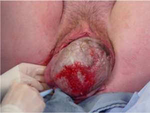
Vaginal Delivery Video Delivery is also known as the second stage of labor, or part of the second stage of labor. It begins with complete dilatation and ends when the baby is completely out of the mother. The exact time of delivery is normally taken at the moment the baby's anterior shoulder (the shoulder delivering closest to the mother's pubic bone) is out. As the fetal head passes through the birth canal, it normally demonstrates, in sequence, the "cardinal movements of labor." These include:
As the fetal head descends below 0 station, the mother will perceive a sensation of pressure in the rectal area, similar to the sensation of an imminent bowel movement. At this time she will feel the urge to bear down, holding her breath and performing a Valsalva, to try to expel the baby. This is called "pushing." The maternal pushing efforts assist in speeding the delivery. For women having their first baby, the second stage will typically take an hour or two. To watch the video, click here Delivery of the baby As the fetal head delivers, support the perineum to reduce the risk of perineal laceration from uncontrolled, rapid delivery. After the fetal head delivers, allow time for the fetal shoulders to rotate and descend through the birth canal. This pause also allows the birth canal to squeeze the fetal chest, forcing amniotic fluid out of the baby's nose and mouth. After a reasonable pause (15-30 seconds), have the woman bear down again, delivering the shoulders and torso of the baby. Clamp and Cut the
Umbilical Cord Once the baby is breathing, put two clamps on the umbilical cord, about an inch (3 cm) from the baby's abdomen. Use scissors to cut between the clamps.
Delivery the Placenta Signs that the placenta is beginning to separate include:
Immediately after the delivery of the baby, uterine contractions stop and labor pains go away. As the placenta separates, the woman will again feel painful uterine cramps. As the placenta descends through the birth canal, she will again feel the urge to bear down and will push out the placenta. If the placenta is not promptly expelled, or if the patient hemorrhages while awaiting delivery of the placenta, this is called a "retained placenta" and it should be manually removed. After delivery of the placenta, the uterus normally contracts firmly, closing off the open blood vessels which previously supplied the placenta. Without this contraction, rapid blood loss would likely prove very problematic or worse. To encourage the uterus to firmly contract, oxytocin 10 mIU IM can be given after delivery. Alternatively, oxytocin 10 or 20 units in a liter of IV fluids can be run briskly (150 cc/hour) into a vein. Breast feeding the baby or providing nipple stimulation (rolling the nipple between thumb and forefinger) will cause the mother's pituitary gland to release oxytocin internally, causing similar, but usually milder effects. A simple way to encourage firm uterine contraction is with uterine massage. The fundus of the uterus (top portion) is vigorously massaged to keep it the consistency of a tightened thigh muscle. If it is flabby, the patient will likely continue to bleed. |
|
|
This information is provided by The Brookside Associates, a private organization, not affiliated with any governmental agency. The opinions presented here are those of the author and do not necessarily represent the opinions of the Brookside Associates. For educational simplicity, only one method is usually shown, but many alternative methods may give satisfactory or superior results. This information is provided solely for educational purposes. The practice of medicine and surgery is regulated by statute and restricted to licensed professionals and those in training under supervision. Performing these procedures outside of that setting is a bad idea, is not recommended, and may be illegal. The presence of any advertising on these pages does not constitute an endorsement of that product or service by the Brookside Associates. C. 2010 All Rights Reserved |
||
 This is the Archived Desktop Edition.