Contents · Introduction · Learning Objectives · Textbook · Lectures · Procedures · Final Exam · Library · Laboratory · Pharmacy · Imaging · Forms · Videos · About · Contact Us>
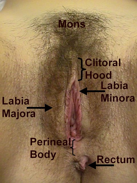
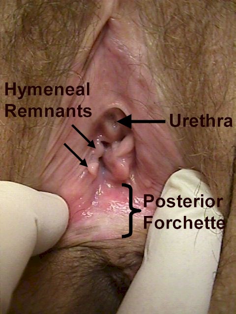
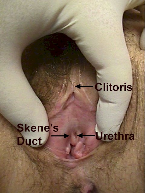
Clinical Anatomy of the VulvaThe vulva is a portal for a variety of functions (reproductive and excretory) and has a unique role in sexual feelings and function. Because it is covered with both dry, squamous skin and moist mucous membrane, it is subject to diseases affecting both. Because of the close proximity of the rectum, intestinal bacteria (anaerobes and coliforms) are more or less constantly present to some degree. These may influence the type and course of infections in this area. The vulva may develop conditions both benign and malignant, symptomless, annoying, or even disabling. The larger lips, labia majora, extend from the mons pubis to the rectum. These are large, fleshy pads that cover the bony pubic rami. They each contain a Bartholin gland, which is usually not noticed but occasionally causes some problems. Just inside the labia majora are the smaller lips, the labia minora. In women who have not had a baby, they are very thin and are usually hidden, to some extent, by the labia majora. After a pregnancy, they are thicker and more prominent. They are rich in nerve endings and are usually very sensitive to touch. During sexual arousal, they swell and moisten with extracellular fluid. During urination, the labia minora function to direct the urine stream in a more or less single direction by forming a curtain on either side of the urethra. The labia minora come together at the top of the vulva to form the clitoral hood. This tissue covers the clitoris, which lies just beneath the hood. The clitoris is characteristically firmer than the surrounding tissues, with a rubbery consistency. It has a high concentration of nerve endings and is extremely sensitive to touch and vibration. It is usually, but not always, the area of greatest sexual sensitivity. During the early stages of sexual arousal, it swells and protrudes just beyond the clitoral hood. In the latter phases of arousal, it generally flattens and retracts back beneath the clitoral hood. The urethra lies between the clitoris and the vagina. It conducts urine from the bladder to the outside. It is normally non-tender to light or moderate touch. On each side of the urethra are the pin-point Skene's ducts. When the labia are spread open, the hymen or remnants of the hymen are visualized. This ring-like structure is usually torn at first intercourse, leading to a small amount of bleeding. For some women, insertion of tampons or other objects will lead to hymeneal rupture. After healing, only small bumps or flaps of skin remain. Anatomic variation with hymens is considerable. Some are thin, stretchy, and so small as to be largely unnoticed. Others are thick and nearly impenetrable. Rarely, the hymen completely covers the vaginal opening, disallowing the passage of menstrual products. Just inside the hymen is the beginning of the vagina. In contrast to the smooth vulvar skin, the vaginal skin has circumferential ridges (rugae). The vagina is not cylindrical in shape, but more like a flattened cone, narrow at the vaginal opening, and widening as the vagina approaches the cervix. Normally, the anterior and posterior vaginal walls are in contact with each other, but during intercourse or an examination, they separate. During sexual arousal, the vaginal skin "sweats" small droplets of extracellular fluid, which is the primary source of lubrication during intercourse. The posterior fourchette is the area of mucous membrane between the rectum and the hymeneal ring. Some women do not seem to have any noticeable discharge, while others normally have a more or less constant slight discharge that is odorless, clear to white and mucoid in nature. |
This information is provided by The Brookside Associates. The Brookside Associates, LLC. is a private organization, not affiliated with any governmental agency. The opinions presented here are those of the author and do not necessarily represent the opinions of the Brookside Associates or the Department of Defense. The presence of any advertising on these pages does not constitute an endorsement of that product or service by either the US Department of Defense or the Brookside Associates. All material presented here is unclassified.
C. 2009, 2014, All Rights Reserved


