LESSON 1 Review of the Circulatory and Respiratory Systems.
TEXT ASSIGNMENT Paragraphs 1-1 through 1-6.
LESSON OBJECTIVES After completing this lesson, you should be able to:
1-1. Identify the general functions of the circulatory system.
1-2. Identify the components of the circulatory system and their functions.
1-3. Identify the general functions of the respiratory system.
1-4. Identify the components of the respiratory system and their functions.
SUGGESTION After you have completed the text assignment, work the exercises at the end of this lesson before beginning the next lesson. These exercises will help you achieve the lesson objectives.
1-1. DEFINITIONS
Some of the terms used in this subcourse are defined below.
a. Casualty. The casualty is the person with the medical problem, such as a person who is not breathing. When being treated by medical personnel, the casualty may be referred to as a patient.
b. Rescuer. The rescuer is the person who is assisting the casualty; for example, the person giving mouth-to-mouth resuscitation to a casualty who is not breathing. In this subcourse, you are the rescuer.
c. Airway. The airway consists of the body structures through which air from the atmosphere passes while going to the lungs.
d. Sign. A sign is anything that the rescuer can tell about the casualty’s condition by using his (the rescuer’s) own senses. For example, a rescuer can see the casualty’s chest rise and fall, hear the sounds made by a casualty when he breathes, and feel the casualty’s pulse.
e. Symptom. A symptom is any change from the norm which is felt by the casualty but which cannot be directly or objectively sensed by the rescuer. Examples of symptoms felt by the casualty include chest pain, nausea, and headache. An injury can produce both signs and symptoms. If you bump your leg against a chair, for example, a bruise may develop. The bruise is a sign of the injury since other people can see the bruise. The pain you feel is a symptom since other people cannot feel your pain.
1-2. IMPORTANCE OF THE CIRCULATORY SYSTEM
The human body is composed of cells. The average adult human’s body is made up of around eighty trillion (80,000,000,000,000) living cells. Cells need energy to survive, repair themselves, perform their functions, and reproduce. Cells obtain this energy through cellular respiration; that is, they combine a source of potential energy with oxygen to liberate energy. The sources of potential energy come from the food (carbohydrates, fats, and proteins) that are processed into usable units by the body’s digestive system (stomach, small intestine, liver, pancreas, and so forth).
The oxygen comes from the air that is inhaled by the lungs. Oxygen in the lungs and food in the intestine cannot help the muscles and other cells unless the oxygen and food can be delivered to those cells. Delivering oxygen and food to the cells is the function of the blood in the body’s circulatory system. The circulatory system also takes waste products (by-products of cellular respiration) from the cells and delivers them to organs (lungs and kidneys) where the wastes can be expelled from the body.
1-3. THE CIRCULATORY SYSTEM
The circulatory system consists of the heart, blood vessels, and blood. The circulatory system brings oxygen and nutrients to the body’s cells and carries away waste products. The circulatory system is also called the cardiovascular system (“cardio-” means heart; “-vascular” means vessels.)
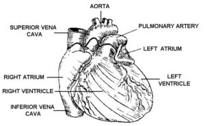
a. Heart. The heart (figure 1-1) is a strong, muscular organ that, by its rhythmic contractions, acts as a force pump maintaining blood circulation. The heart is about the size of a fist and is located in the lower left-central part of the chest cavity.

(1) Layers. The heart consists of three layers.
(a) The myocardium is the middle layer. It is composed of the actual heart muscles. (“Myo-” means muscle; “cardium” means heart.)
(b) The pericardium is the outer layer. It is a double-walled sac that surrounds the heart muscles. (“Peri-” means around.)
(c) The endocardium is the inner layer. It forms the inner lining of the four chambers. (“Endo-” means within.)
(2) Chambers. The heart can be described as being two pumps. Each side (right half and left half) of the heart has a receiving chamber for the blood (the atrium) and a pumping chamber (the ventricle). The two halves of the heart are separated by a wall-like structure called the interventricular septum.
NOTE: The plural of atrium is atria.
(3) Sinoatrial node. The sinoatrial (SA) node is a small bundle of nerve tissue located at the junction of the superior vena cava and the right atrium. The sinoatrial node is a natural pacemaker that produces an electrical stimulus. This electrical stimulus causes the muscles of the ventricles to contract and pump blood.
b. Blood Vessels. The blood vessels are firm, elastic, muscular tubes that carry the blood away from the heart and back to the heart again.
(1) Blood circulation systems. Since the heart is divided into two parts (the right half consisting of the right atrium and the right ventricle and the left half consisting of the left atrium and left ventricle), it is not surprising to find that there are actually two blood circulatory systems–the systemic and the pulmonary.
(a) Systemic. The systemic (general) circulatory system is the larger of the two systems. It takes the blood pumped by the left ventricle to all parts of the body and returns the blood to the right atrium. The oxygen content of the blood is high when it leaves the heart through the left ventricle and is low when it returns to the right atrium.
(b) Pulmonary. The pulmonary circulatory system takes the blood pumped by the right ventricle to the lungs and returns the blood to the left atrium. The oxygen content of the blood is low when it leaves the heart through the right ventricle and high when it returns to the left atrium.
(2) Types of blood vessels. Both the systemic and the pulmonary circulatory systems are composed of three major types of blood vessels–arteries, capillaries, and veins.
(a) Arteries. The arteries carry blood pumped by the ventricles away from the heart. The arteries of the systemic circulatory system carry oxygenated (oxygen rich) blood to body tissues. The pulmonary arteries carry deoxygenated (oxygen-poor) blood to the lungs. Arteries have the capacity to constrict and dilate. This constricting and dilating helps to regulate the blood pressure.
(b) Capillaries. Originally, the arteries are large blood vessels. Soon, however, they divide into smaller branches. These branches then divide again and again. With each division, the blood vessels become smaller and smaller. Finally, the blood vessels are so small that only one red blood cell can pass through at a time. When they reach this size, the blood vessels are called capillaries. When a red blood cell enters the capillaries, it is free to perform its primary functions.
In the pulmonary system, red blood cells give up carbon dioxide to the lungs and pick up oxygen. In the systemic system, red blood cells give oxygen and nutrients to the cells and pick up carbon dioxide and other waste products.
(c) Veins. Capillaries join together to form larger blood vessels, which then combine to form even larger blood vessels. These blood vessels are called veins. Veins carry the blood back to the heart. The veins of the systemic system carry oxygen-poor blood to the right atrium. The veins of the pulmonary system carry oxygen-rich blood to the left atrium. The veins are not as thick as the arteries, and they will collapse when severed. Many veins have valves, which keep blood from flowing backward (away from the heart). The term “vena” denotes a vein.
c. Blood. Blood is a viscous (thick), reddish fluid. When the blood is oxygenated (oxygen-rich), it is bright red. When the blood is low in oxygen content, it is a darker red. When the darker color is seen through a layer of skin tissue, it appears to be bluish. Blood is composed of fluid and solids.
(1) Plasma. The liquid part of the blood is called plasma. It is straw-colored (pale yellow) and carries the solid components of the blood such as erythrocytes, leukocytes, and thrombocytes.
(2) Erythrocytes. Erythrocytes (also called red blood cells or RBC) transport oxygen from the lungs and nutrients from the small intestine to the cells of the body. They also transport carbon dioxide and other waste materials from the body’s cells to the lungs and kidneys where the waste products are removed and expelled.
(3) Leukocytes. Leukocytes (also called white blood cells or WBC) assist in the body’s defense against disease by attacking and destroying bacteria and other foreign particles in the blood and body tissues.
(4) Thrombocytes. Thrombocytes (also called platelets) help to stop bleeding from a damaged blood vessel. Although thrombocytes normally show no tendency to coagulate (clot) in the blood, they change character when they approach a cut or tear in a blood vessel. The thrombocytes then combine to form a soft clot where the vessel wall is broken. This clot soon hardens to form a plug to stop the loss of blood.
1-4. BLOOD FLOW
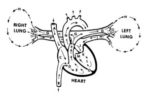
In order to summarize how blood flows in the body, let’s take a trip through the body’s circulatory system (figure 1-2). We will enter the system at the vena cava.
a. Vena Cava. There are two major blood veins, which empty into the right atrium. The superior vena cava carries oxygen-poor blood coming from the head, arms, and chest. The inferior vena cava returns oxygen-poor blood from the lower trunk and legs.

|
Figure 1 Labels |
|
| A. Superior vena cava | I. Pulmonary veins |
| B. Inferior vena cava | J. Left atrium |
| C. Right atrium | K. Mitral valve |
| D. Tricuspid valve | L. Left ventricle |
| E. Right ventricle | M. Aortic valve |
| F. Pulmonary valve | N. Aorta |
| G. Pulmonary arteries | O. Interventricular septum |
| H. Lungs | |
b. Right Atrium. The right atrium receives blood from the superior vena cava and the inferior vena cava. When the right ventricle relaxes (that is, after it has contracted and pumped blood), blood flows from the right atrium into the right ventricle through the tricuspid valve. The tricuspid valve is formed so that blood cannot flow back into the right atrium when the right ventricle contracts.
c. Right Ventricle. When the right ventricle is filled with blood, it receives an impulse from the sinoatrial node. This impulse causes the muscles of the right ventricle to contract. This contraction causes the inside of the ventricle (the space where the blood is) to become smaller. The increased pressure forces blood out of the ventricle and into the pulmonary artery. The pulmonary valve located at the beginning of the pulmonary artery keeps blood from flowing back into the right ventricle when the ventricle relaxes and returns to its normal size.
d. Lungs (Pulmonary System). The pulmonary artery divides into two arteries. One artery travels to the right lung while the other artery travels to the left lung. The arteries divide until they reach the capillary stage. The capillaries surround the alveoli (air sacs) of the lungs. There the oxygen-poor blood gets rid of carbon dioxide and picks up oxygen from the air in the alveolus. The blood, now high in oxygen content, then returns to the left atrium through the pulmonary veins.
e. Left Atrium. The left atrium receives blood from the lungs through two pulmonary veins. When the left ventricle relaxes after having contracted, the blood flows from the left atrium into the left ventricle through the mitral valve. The mitral valve keeps blood from flowing back into the left atrium when the left ventricle contracts.
f. Left Ventricle. After the left ventricle is filled with oxygen-rich blood, it receives an impulse from the sinoatrial node, which causes it to contract and pump blood into the large artery call the aorta. When the blood enters the aorta, it passes through the aortic valve. This valve keeps the blood from flowing back into the heart once the left ventricle relaxes.
g. Body (Systemic System).
(1) Some arteries branch off the aorta to provide the brain, upper body, and heart with blood. Blood returns to the heart from these areas through the superior vena cava.
NOTE: If blood flow to the brain stops and is not restored (either the casualty’s heart starts beating on its own or cardiopulmonary resuscitation is administered), the brain will begin to die in six to ten minutes.
(2) The aorta turns down and divides into smaller arteries which go to the lower parts of the body. Some of the blood picks up fluids and nutrients from the intestines. Some of the blood passes through the liver and kidneys which remove bacteria and other unwanted substances from the blood. The blood returns to the heart from these areas through the inferior vena cava.
1-5. THE RESPIRATORY SYSTEM
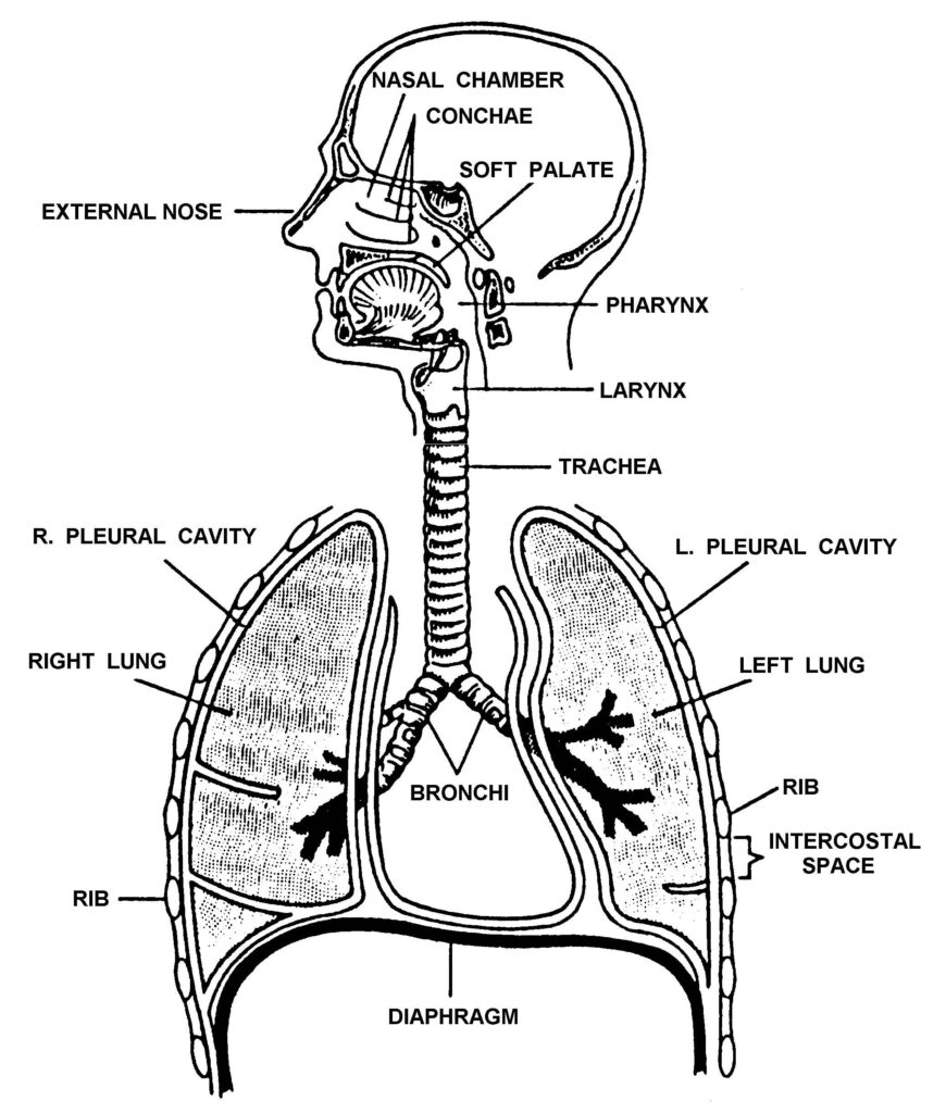
The respiratory system consists of two lungs and the respiratory tract that carries air to and from the lungs (figure 1-3). When a person inhales, the air enters the nose or mouth, travels down the trachea, and into the two bronchi. Each bronchus divides into smaller and smaller air tubes. Finally, the air reaches the alveoli (air sacs). The red blood cells in the capillaries surrounding the alveoli absorb oxygen from the air and give off carbon dioxide, which passes into the alveoli.
When a person exhales, the air travels from the alveoli through the air tubes, up the trachea, and out the nose or mouth. Of course, not all of the air inhaled reaches the alveoli nor is all of the oxygen removed from the air in the alveoli. The average adult takes in about 500 milliliters (ml) of air each time he inhales and he exhales the same amount. Even after the person exhales, the lungs still contain about 2300 ml of air. The anatomy (structures) and the physiology (functions) of the respiratory system are briefly discussed below.
a. Nose. The nose is composed of two nostrils (openings) and two nasal cavities (air chambers above the roof of the mouth and below the cranium). A structure called the nasal septum separates the right nostril and nasal cavity from the left nostril and nasal cavity. The nose warms, moistens, and filters the inhaled air. Special nerve endings in the upper part of the nasal cavities provide the sense of smell.
b. Pharynx. The pharynx is a part of the throat that is part of both the respiratory system and the digestive system. The pharynx is divided into three parts. The nasopharynx (upper part) connects with the nasal chambers. The oropharynx (middle part) connects with the oral cavity (mouth). The laryngopharynx (lower part) connects with the larynx (respiratory system) and the esophagus (digestive system).
c. Epiglottis. The epiglottis is a flap that covers the entrance to the larynx when a person swallows. This prevents food from entering the larynx instead of the esophagus. When a person inhales, the entrance to the larynx is not covered and air enters the larynx. If a foreign object enters the airway, it can block the airway and cause breathing to stop.
d. Larynx. The larynx is a box-like structure composed of cartilage, ligaments, and muscles that sits on top of the trachea. The larynx contains the vocal cords, which produce the voice; therefore, it is sometimes called the voice box. It is also called the Adam’s apple because of the bulge it causes in the throat.

e. Trachea. The trachea (windpipe) is a tube composed of horseshoe-shaped rings of cartilage. Cilia (hair-like projections) on the inner lining of the trachea help to filter air as it passes through the trachea to the bronchi.
f. Bronchi. The bronchi are two tube-like structures at the base of the trachea. One bronchus leads toward the right lung; the other bronchus leads toward the left lung. Like the trachea, the bronchi are composed of cartilaginous rings and are lined with a mucous membrane.
g. Bronchioli. The bronchi divide and subdivide until they become small air tubes one millimeter or less in diameter call bronchioli. The bronchioli continue to subdivide until they become very small tubes ending in alveoli.
h. Alveoli. Alveoli are tiny, grape-like clusters of microscopic air sacs. Air enters the alveoli from the bronchioli. The wall of an alveolus is one cell layer thick. The alveolus is surrounded by equally thin capillaries. Oxygen (O2) molecules from the air inside the alveolus travel through the alveolus and capillary walls to the blood within the capillary. The hemoglobin in the red blood cells captures the oxygen molecules and release carbon dioxide (CO2) molecules. The carbon dioxide molecules and some water molecules travel from the blood, through the walls, and into the alveoli.
i. Lungs. The alveoli, bronchioli, and associated blood vessels make up two cone-like organs called lungs. The lungs are broad at their base (which rests on the diaphragm) and narrow at the apex (top). Each lung is surrounded by pleural membranes that prevent friction when the lung expands and contracts. The right lung is divided into three lobes; the left lung is divided into two lobes. The left lung is smaller than the right lung because the heart takes up space on the left side of the chest cavity.
1-6. MECHANICS OF BREATHING
Breathing refers to the process of moving air into and out of the lungs. The process is usually performed automatically (without conscious thought) by the respiratory control center located in the medulla oblongata of the brain stem. The normal range of breathing rates (one cycle consists of one inspiration and one exhalation) in an adult is 12 to 20 breaths per minute. Regular, easy breathing is referred to as eupnea. Difficulty in breathing is referred to as dyspnea.
a. Inhalation. During the inhalation (inspiration) phase of breathing, the diaphragm and the intercostal muscles contract. When the diaphragm muscle (located at the base of the lungs) contracts, it is pulled downward toward the abdomen. This flattening of the diaphragm enlarges the chest cavity. When the intercostal muscles (located between the ribs) contract, they lift the rib cage up and out (chest rises). This also enlarges the chest cavity. This expansion of the chest cavity causes the air pressure in the alveoli to decrease. Air from the outside environment rushes in through the nose or mouth to equalize the pressure.
b. Exhalation. During the exhalation (expiration) phase of breathing, the diaphragm and the intercostal muscles relax. When the diaphragm muscle relaxes, it resumes its dome-like shape (moves upward). When the intercostal muscles relax, they let the rib cage return to its original position (moves down and inward). Both of these actions cause the air pressure in the alveoli to increase and force air out of the lungs, through the airways, and out the nose or mouth.
