|
Medical Education Division |
Operational Medicine 2001
Standard First Aid Course
NAVEDTRA 13119


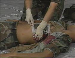
BleedingBleeding (hemorrhage) is the escape of blood from capillaries, veins, and arteries. Capillaries are very small blood vessels that carry blood to all parts of the body. Veins are blood vessels that carry blood to the heart. Arteries are large blood vessels that carry blood away from the heart. Bleeding can occur inside the body (internal), outside the body (external) or both. Blood is a fluid that consists of a pale yellow liquid (plasma), red blood cells (erythrocytes), white blood cells (leukocytes), and platelets (thrombocytes). Plasma is the fluid portion of the blood that carries nutrients. Red blood cells give color to the blood and carry oxygen. White blood cells defend the body against infection and attack foreign particles. Platelets are disk shaped and assist in clotting the blood, the mechanism that stops bleeding. There are three types of bleeding. Capillary bleeding is slow, the blood "oozes" from the (wound) cut. Venous bleeding is dark red or maroon, the blood flows in a steady stream. Arterial bleeding is bright red, the blood "spurts" from the wound. Arterial bleeding is life threatening and difficult to control. In small wounds, only the capillaries are damaged. Deeper wounds result in damage to the veins and arteries. Damage to the capillaries is usually not serious and can easily be controlled with a Band-Aid. Damage to the veins and arteries are more serious and can be life threatening. The adult body contains approximately 5 to 6 quarts of blood (10 to 12 pints). The body can normally lose 1 pint of blood (usual amount given by donors) without harmful effects. A loss of 2 pints may cause shock, a loss of 5 to 6 pints usually results in death. During certain situations it will be difficult to decide whether the bleeding is arterial or venous. The distinction is not important. The most important thing to remember is that all bleeding must be controlled as soon as possible. External Bleeding While administering first aid to a casualty who is bleeding, you must remain calm. The sight of blood is an emotional event for many, and it often appears severe. However, most bleeding is less severe than it appears. Most of the major arteries are deep and well protected by tissue and bone. Although bleeding can be fatal, you will usually have enough time to think and act calmly. There are four methods to control bleeding: direct pressure, elevation, indirect pressure, and the use of a tourniquet. Direct Pressure Direct pressure is the first and most effective method to control bleeding. In many cases, bleeding can be controlled by applying pressure directly (Fig. 3-1) to the wound. Place a sterile dressing or clean cloth on the wound, tie a knot or adhere tape directly over the wound, only tight enough to control bleeding. If bleeding is not controlled, apply another dressing over the first or apply direct pressure with your hand or fingers over the wound. Direct pressure can be applied by the casualty or a bystander. Under no circumstances is a dressing removed once it has been applied. Elevation Raising (elevation) of an injured arm or leg (extremity) above the level of the heart will help control bleeding.
Figure 3-1 Direct Pressure
Figure 3-2 Pressure Points for Control of Bleeding Elevation should be used together with direct pressure. Do not elevate an extremity if you suspect a broken bone (fracture) until it has been properly splinted and you are certain that elevation will not cause further injury. Use a stable object to maintain elevation. Placing an extremity on an unstable object may cause further injury. Indirect Pressure In cases of severe bleeding when direct pressure and elevation are not controlling the bleeding, indirect pressure must be used. Bleeding from an artery can be controlled by applying pressure to the appropriate pressure point. Pressure points (Fig. 3-2) are areas of the body where the blood flow can be controlled by pressing the artery against an underlying bone. Pressure is applied with the fingers, thumb, or heel of the hand. Pressure points should be used with caution. Indirect pressure can cause damage to the extremity due to inadequate blood flow. Do not apply pressure to the neck (carotid) pressure points, it can cause cardiac arrest. Indirect pressure is used in addition to direct pressure and elevation. Pressure points in the arm (brachial) and in the groin (femoral) are most often used, and should be thoroughly understood. The brachial artery is used to control severe bleeding of the lower part of the upper arm and elbow. It is located above the elbow on the inside of the arm in the groove between the muscles. Using your fingers or thumb, apply pressure (Fig. 3-2E) to the inside of the arm over the bone. The femoral artery is used to control severe bleeding of the thigh and lower leg. It is located on the front, center part of the crease in the groin. Position the casualty on his or her back, kneel on the opposite side (Fig. 3-2H ) from the wounded leg, place the heel of your hand directly on the pressure point, and lean forward to apply pressure. If the bleeding is not controlled, it may be necessary to press directly over the artery with the flat surface of the fingertips and to apply additional pressure on the fingertips with the heel of your other hand. A tourniquet should be used only as a last resort to control severe bleeding after all other methods have failed and is used only on the extremities. Before use, you must thoroughly understand its dangers and limitations. Tourniquets cause tissue damage and loss of extremities when used by untrained individuals. Tourniquets are rarely required and should only be used when an arm or leg has been partially or completely severed and when bleeding is uncontrollable. The standard tourniquet is normally a piece of cloth folded until it is 3 or more inches wide and 6 or 7 layers thick. A tourniquet can be a strap, belt, neckerchief, towel, or other similar item. A folded triangular bandage makes a great tourniquet. Never use wire, cord, or any material that will cut the skin. To apply a tourniquet (Fig. 3-3), do the following: 1. While maintaining the proper pressure point, place the tourniquet between the heart and the wound, leaving at least 2 inches of uninjured skin between the tourniquet and wound. 2. Place a pad (roll) over the artery. 3. Wrap the tourniquet around the extremity twice, and tie a half-knot on the upper surface. 4. Place a short stick or similar object on the half-knot, and tie a square knot. 5. Twist the stick to tighten, until bleeding is controlled. 6. Secure the stick in place. 7. Never cover a tourniquet.
Figure 3-3 Applying a Tourniquet 8. Using lipstick or marker, make a 'T" on the casualty's forehead and the time tourniquet was applied. 9. Never loosen or remove a tourniquet once it has been applied. The loosening of a tourniquet may dislodge clots and result in enough blood loss to cause shock and death. Do not touch open wounds with your fingers unless absolutely necessary. Place a barrier between you and the casualty's blood or body fluids, using plastic wrap, gloves, or a clean, folded cloth. Wash your hands with soap and warm water immediately after providing care, even if you wore gloves or used another barrier. Internal Bleeding Internal bleeding, although not usually visible, can result in serious blood loss. A casualty with internal bleeding can develop shock before you realize the extent of their injuries. Bleeding from the mouth, ears, nose, rectum, or other body opening (orifice) is considered serious and normally indicates internal bleeding. The most common sign of internal bleeding is a simple bruise (contusion), it indicates bleeding into the skin (soft tissues). Severe internal bleeding occurs in injuries caused by a violent force (automobile accident), puncture wounds (knife), and broken bones. Signs of internal bleeding include: 1. Anxiety and restlessness. 2. Excessive thirst (polydipsia). 3. Nausea and vomiting. 4. Cool, moist, and pale skin (cold and clammy). 5. Rapid breathing (tachypnea). 6. Rapid, weak pulse (tachycardia). 7. Bruising or discoloration at site of injury (contusion). If you suspect internal bleeding, do the following: 1. Bruise (contusion) - Apply ice or cold pack, with cloth to prevent damage to the skin, to reduce pain and (edema) swelling. 2. Severe internal bleeding:
Nosebleeds (epistaxis) can be caused by an injury, disease, the environment, high blood pressure, and changes in altitude. They frighten the casualty and may bleed enough to cause shock. If a fractured skull is suspected as the cause, do not stop the bleeding. Cover the nose with a loose, dry, sterile dressing and call the local emergency number or medical personnel. If the casualty has a nosebleed due to other causes, do the following: 1. Keep the casualty quiet, sitting with head tilted forward. 2. Pinch the nose shut (if there is no fracture), place ice or cold packs to the bridge of the nose, or put pressure on the upper lip just below the nose. Inform the casualty not to rub, blow, or pick his or her nose. Seek medical assistance if the nosebleed continues, bleeding starts again, or bleeding is because of high blood pressure. If the casualty loses consciousness, place them on their side to allow blood to drain from the nose and call the local emergency number or medical personnel. Foreign bodies in the nose usually occur among children. First aid consists of seeking professional medical attention. Nasal damage and the possibility of pushing the object farther up the nose can result from searching and attempts at removal by unqualified personnel. Casualties with severe external bleeding and suspected internal bleeding must be seen by medical personnel as soon as possible. All casualties with external and internal bleeding should be treated for shock. References 1. Karren, K. J. and Hafen, B.Q.: First Responder A Skills Approach, edition 3, Morton
Publishing Company
Department of the Navy Approved for public release; Distribution is unlimited. The listing of any non-Federal product in this CD is not an endorsement of the product itself, but simply an acknowledgement of the source. Operational Medicine 2001 Home · Military Medicine · Sick Call · Basic Exams · Medical Procedures · Lab and X-ray · The Pharmacy · The Library · Equipment · Patient Transport · Medical Force Protection · Operational Safety · Operational Settings · Special Operations · Humanitarian Missions · Instructions/Orders · Other Agencies · Video Gallery · Phone Consultation · Forms · Web Links · Acknowledgements · Help · Feedback
*This web version is provided by The Brookside Associates Medical Education Division. It contains original contents from the official US Navy NAVMED P-5139, but has been reformatted for web access and includes advertising and links that were not present in the original version. This web version has not been approved by the Department of the Navy or the Department of Defense. The presence of any advertising on these pages does not constitute an endorsement of that product or service by either the US Department of Defense or the Brookside Associates. The Brookside Associates is a private organization, not affiliated with the United States Department of Defense. |


