Hospital Corpsman Sickcall Screener's Handbook
BUMEDINST 6550:9A
Naval Hospital Great Lakes
1999
Thorax, Lungs, and Respiratory Disorders
Allotted time:
|
 |
|
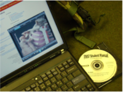
Operational Medicine CD
Text, images,
videos and manuals
The essential text for military healthcare providers
www.brooksidepress.org |
Instructional references:
Instructional aids:
-
visual aid panel
-
transparencies
-
student handout
Terminal learning objective: Given a simulated patient with simulated symptoms, the student will be able to recognize and correctly examine the patient using proper procedure.
Enabling learning objective:
-
Identify proper land marks of the thorax and lungs. -
Properly inspect, palpate, percuss, and auscultate. -
Identify breath sounds. -
Identify the different respiratory disorders. -
Be able to differentiate between the different types of pneumonia. -
Identify the signs and symptoms of common respiratory disorders. -
Identify the treatment of common respiratory conditions.
-
Landmarks - anatomical structures
-
Anterior
-
mid sternal line - vertical - down center of sternum.
-
right and left midclavicular line - midpoint of clavicle.
-
right and left anterior axillary line.
-
suprasternal notch - top of sternum.
-
sternal angle - where the manubrium and sternum meet.
-
xiphoid process - distal to sternum.
-
Lateral
-
right and left anterior axillary line.
-
mid axillary - vertical from apex of axilla.
-
posterior axillary line - vertical from posterior axillary fold.
-
Posterior
-
right and left posterior axillary lines.
-
right and left scapula line - vertical from inferior angle of scapula.
-
vertebral line - vertical along spinous processes.
-
Lungs
-
apex 2-4 cm above inner one-third of clavicle
-
inferior anterior border - crosses 6th rib at midclavicular line and 8th rib at mid axillary line.
-
inferior posterior border - at level of 10th thoracic spinous process (T-12 at deep inspiration)
-
Tracheal bifurcation - left and right mainstem bronchus at sternal angle (anterior) and T-4 (posterior)
-
Five lobes of the lungs - left upper lobe (LUL), left lower lobe (LLL), right upper lobe (RUL), right middle lobe (RML), and right lower lobe (RLL). These will vary in position and size during phases of respiration.
-
Lingula is part of the lung that lies adjacent to the heart.
-
Exam Techniques
-
General approach
-
Thorax exposed in good lighting, undressed to waist.
-
Proceed in order - inspect, palpate, percuss, and auscultate.
-
Try to visualize underlying tissue.
-
Survey of thorax and respiration
-
Patients color
-
Shape of fingernails
-
Position of trachea
-
Respiratory distress
-
Observe rate and rhythm and effort of breathing
-
Inspect next for supraclavicular retractions or sternocleidomastoid contractions
-
Listen to breathing
-
Observe shape of chest
-
Exam
-
Inspection
-
Deformities or asymmetries
-
kyphosis (hunchback)
-
lordosis (backward curvature of spine)
-
scoliosis (s-shaped lateral curvature of spine)
-
pectus carinatum (pigeon chest)
-
pectus excauatum (caved in chest)
-
barrel chest (increased anterior - posterior diameter)
-
slope of ribs
-
more horizontal in emphysema, severe asthma or airway obstruction.
-
intercostal retractions during inspirations
-
severe asthma, emphysema or laryngeal/tracheal obstruction
-
local bag or impaired respiratory motion - underlying pleural or lung disease.
-
Palpation of the chest
-
Uses
-
identifies areas of tenderness
-
assessment of observed abnormalities
-
assessment of respiratory excursion - (lag or impaired inspiration)
-
Technique
-
place thumb level/parallel to 10th ribs bilaterally
-
grasp lateral rib cage with hands
-
patient inhales deeply
-
watch movement of thumbs
-
Technique to elicit vocal or tactile fremitus
-
fremitus refers to palpable vibrations transmitting through the chest wall.
-
technique
-
use ball of hand
-
ask patient to repeat the words "blue moon", "one on one", or "ninety-nine".
-
palpate and compare symmetrical areas
-
Fremitus decreased in
-
bronchial obstruction
-
soft voice intensity
-
pleural space disease
-
pneumo thorax
-
COPD
-
infiltrating tumor
-
very thick chest wall
-
Fremitus increased
-
near large bronchi
-
over consolidated lung
-
Identification of level of diaphragm
-
using ulnar side of hand, place at expected level
-
move hand up and down until fremitus no longer felt
-
this approximates level of the diaphragm
-
Percussion of the chest
-
General principles
-
sets wall/underlying tissue in motion
-
produces audible sounds/palpable vibrations
-
aids in determining if underlying tissue is:
-
air filled
-
fluid-filled
-
solid
-
Penetrates approximately 5-7 cm into chest.
-
Technique
-
Hyperextend middle finger and place distal phalanx and D.I.P. joint firmly on surface to be percussed. Avoid contact with other part of hand. -
Partially flex the middle finger (plexor) of the other hand with hand locked upwards. -
Strike pleximeter finger at the base of the distal phalanx quick and sharp with the plexor movement should be at the wrist not finger. -
Remove striking finger quickly.
-
Strike 2-3 times in each exam area.
-
Compare one part of chest with opposite side.
-
Five basic percussion notes
-
Flatness
-
soft intensity
-
high pitch
-
short duration
-
example/location-thigh
-
seen with large pleural effusion
-
Dullness
-
medium intensity
-
medium pitch
-
medium duration
-
example/location - liver
-
seen with lobar pneumonia
-
Resonance
-
loud intensity
-
low pitch
-
long duration
-
example/location - normal lung
-
bronchitis
-
Hyperresonance
-
very loud
-
low pitch
-
clonger duration
-
example/location - normally none
-
emphysematous lung, pneumothorax
-
Tympany
-
loud
-
high pitch
-
variable duration
-
example/location - gastric air bubble
-
large pneumothorax
-
Areas to percuss
-
across top of each shoulder
-
downward in intervals between scapulas to level of diaphragm
-
areas lateral to mid-scapular lines
-
describe abnormal percussion
-
identify diaphragmatic level
-
Auscultation of the chest
-
Principles of exam
-
use diaphragm of stethoscope
-
use same locations as percussion
-
listen to one full breath in each area
-
watch for hyperventilation, faintness and light-headedness
-
auscultate side to side so that right to left comparison is made
-
Auscultate breath sounds
-
Intensity - decreased with shallow breath respirations, thick chest (obesity), COPD, decreased transmission as in pleural effusion or pneumothorax.
-
Pitch and duration of breath sounds.
-
Are sounds during inspiration/expiration, or both?
-
Normal distribution of sounds?
-
Normal sounds in abnormal places?
-
Adventitious sounds - crackles wheezes or rubs.
-
location
-
location in which phase
-
Breath sounds/auscultation - patient always breaths through the mouth.
-
normal breath sounds
-
vesicular sounds
-
low in pitch of expiration
-
soft in intensity
-
normal location throughout most of lungs away from trachea/large bronchi
-
last longer during inspiration
-
Bronchial sounds
-
high in pitch
-
loud in intensity
-
normal location near larger airway
-
expiratory sounds equal or longer
-
Bronchovesicular sounds
-
intermediate pitch
-
intermediate intensity
-
normal location is the 1st and 2nd interspace and between scapula
-
equal on inspiration/expiration
-
Tracheal sounds
-
relatively high in pitch
-
very loud in intensity
-
normal location is over trachea in neck
-
equal on inspiration and expiration
-
Adventitious sounds - note timing in cycles
-
Crackles (or rales)
-
dry or moist crackling sounds
-
may occur during inspiration or both
-
discrete non continuous sounds
-
noted in pneumonia, pulmonary edema, luminary fibrosis
-
Two types
-
fine crackles - soft high pitched
-
coarse crackles - somewhat louder, lower in pitch
-
Rhonchi - coarse, low pitch snoring sounds
-
Wheezes
-
musical, higher pitched, hissing or shrill
-
may be expiratory or inspiratory
-
Pleural rubs
-
loud, rubbing quality
-
localized
-
often inspiratory and expiratory
-
Voice sounds: More valuable in detecting consolidation, infarction, or etelectosis. Normally faint and indistinct except over bronchi.
-
Egophony - pt says "EEE", you hear "Ay". This is due to increased transmission through consolidated or airless lungs.
-
Whispered pectoriloquy - whispered sounds heard more clearly through consolidated lung tissue.
-
Bronchial breath sounds in peripheral areas.
-
Bronchophony - louder, clearer voice sounds because of increased transmission of high pitched components.
-
Exam of anterior chest
-
Inspection
-
rate, rhythm, effort of respirations
-
listen to breathing
-
shape/movement of the chest
-
width of costal angle
-
retraction of interspaces with inspiration
-
local lag/impaired respiratory
-
Palpation
-
Hands are placed along costal margin with fingers lateral along border of rib cage.
-
observe symmetry, range of excursion
-
Tactile/vocal fremitus
-
utilize same technique as described previously
-
compare symmetric areas
-
Percussion
-
Same technique as described previously
-
Auscultation
-
listen to breath sounds
-
note intensity
-
variations of normal breath sounds
-
bronchial breath sounds over large airways
-
added sounds
-
May have your patient breath hard and fast through open mouth.
-
Clinical assessment of pulmonary function
-
Ambulate patient down hall or climb stairs
-
assess complaint
-
Match test
-
Hold lighted match 6 inches from patients mouth and have patient blow out match with open mouth.
-
Inability indicates severe obstruction
-
Abnormalities in rate and rhythm
-
Rapid, shallow breathing (tachypnea)
-
Has numerous causes
-
Rapid, deep breathing (hyperpnea) (hyperventilation)
-
May be due to exercise, anxiety.
-
Slow breathing (bradypnea)
-
diabetic coma, respiratory depression
-
Cheyne-Stokes breathing
-
Alternating periods of deep breathing with periods of apnea
-
May be due to heart failure, respiratory depression
-
Basic diseases of the lower respiratory tract.
-
Basic examination
-
pt should be undressed to waist
-
proceed in order
-
inspection, palpation, percussion and auscultation
-
compare sides
-
work from top to bottom
-
Try to visualize underlying tissues and organs
-
pt sitting: examine posterior thorax and lungs
-
pt standing: examine anterior thorax and lungs
-
Disorders
-
Pneumonia: an acute infection of the alveolar spaces and/or interstitial tissue of the lungs
-
Pneumococcal Pneumonia (streptococcus pneumonia) most common causes of lobar pneumonia.
-
Signs and Symptoms
-
proceeded by URI
-
sudden onset/rapid progression
-
sharp pain in the involved hemi thorax
-
productive cough with yellow green, gray, or rusty colored sputum
-
dyspnea, tachycardia
-
shaking chills, fever
-
pleural friction rubs
-
patient most comfortable lying on affected side
-
rales in affected lobes
-
Diagnosis
-
Should be suspected when:
-
pt exhibits any of above symptoms
-
diagnosis supported by physical exam
-
chest X-ray, CBC, and if possible - sputum C&S
-
Refer to MO if you suspect pneumonia
Other bacterial pneumonias
-
Chlamydia pneumonia - fever, previous URI, non-productive cough, mild to moderate illness, normal
WBC, minimal physical findings. Small segmental infiltrates on
chest X-ray.
-
Haemophilus influenza - patient is moderately ill.
-
Legionella pneumonia - very severe illness. High fever, non-productive cough, chest pain, neurolgic changes, G.I. disturbances.
-
Mycoplasma pneumonia - slow onset, headache, malaise, fever, scratchy sore throat, dry cough. Mild and self limited, will resolve in 2-4 weeks without treatment. X-ray may show patchy infiltrates. Might have some crackles or isolated wheezing.
-
Bronchitis - inflammation of the trachea and bronchial tree.
-
Etiology
-
may develop following a cold or other viral infections
-
exposure to pollutants and other irritants
-
Signs and symptoms
-
proceeded by URI
-
malaise, fever, muscle pain, sore throat
-
cough: initially dry and non-productive followed by sputum which may become abundant and mucopurulant with a greenish - yellowish color.
-
may hear rhonchi, but not rales
-
Diagnosis
-
possible by signs and symptoms
-
do chest X-ray to rule out other complications
-
Treatment
-
rest, increase fluids
-
antipyretics, cough suppressant
-
bronchodilators (when patient is wheezing)
-
antibiotics when sputum or fever
-
Asthma - A bronchial hypersensitivity disorder characterized by reversible airway obstruction.
-
Etiology: Hyperactive airways with attacks of bronchospasms initiated by various factors such as:
-
allergic reactions
-
inhalation of irritants
-
exercise
-
stress
-
infection
-
Signs and symptoms
-
wheezing, musical in nature
-
dyspnea, coughing with sputum
-
night coughing and wheezing on exertion
-
Treatment
-
refer to MO
-
acute attack requires aggressive treatment
-
bronchodialators required for treatment
-
Pleurisy - pain secondary to inflammation of the pleura (pleuritis).
-
Etiology
-
pleural injury
-
entry of infectious agent
-
pleural trauma
-
pulmonary embolism
-
Signs and symptoms
-
sudden onset
-
pain which is increased by coughing and breathing
-
may hear pleural friction rub
-
Diagnosis
-
characteristics pain
-
X-ray to rule out other causes
-
Treatment
-
refer to MO
-
analgesics, bronchodialators and antibiotics
-
treat any other underlying causes
|
|
Approved for public release;
Distribution is unlimited.
The listing of any non-Federal product in this CD is not an endorsement of the
product itself, but simply an acknowledgement of the source.
Bureau of Medicine and Surgery
Department of the Navy
2300 E Street NW
Washington, D.C
20372-5300 |
Operational Medicine
Health Care in Military Settings
CAPT Michael John Hughey, MC, USNR
NAVMED P-5139
January 1, 2001 |
United States Special Operations
Command
7701 Tampa Point Blvd.
MacDill AFB, Florida
33621-5323 |
*This web version is provided by
The Brookside Associates Medical Education Division. It contains
original contents from the official US Navy NAVMED P-5139, but has been
reformatted for web access and includes advertising and links that were not
present in the original version. This web version has not been approved by the
Department of the Navy or the Department of Defense. The presence of any
advertising on these pages does not constitute an endorsement of that product or
service by either the US Department of Defense or the Brookside Associates. The
Brookside Associates is a private organization, not affiliated with the United
States Department of Defense.
Contact Us · Other
Brookside Products

|
|
Operational Medicine 2001
Contents
|
|
 |
|
FMST Student Manual Multimedia CD
30 Operational Medicine Textbooks/Manuals
30 Operational Medicine Videos
"Just in Time" Initial and Refresher Training
Durable Field-Deployable Storage Case |
|