Hospital Corpsman Sickcall Screener's Handbook
BUMEDINST 6550:9A
Naval Hospital Great Lakes
1999
Examination of the Abdominal Region
Terminal learning objective: Given a simulated patient with simulated symptoms, the student will be able to recognize potential problems and properly perform the needed exam.
-
Enabling learning objective:
-
Identify different bowel sounds. -
Identify different types of hernias. -
Identify different organs and their position in the abdominal cavity. -
Identify the different symptoms of an acute abdomen.
-
References:
-
Taber’s Cyclopedic Medical Dictionary, 1989
-
The Merck Manual, Sixteenth Edition.
-
Anatomy & Physiology
-
The abdomen is divided into 4 quandrants.
-
RUQ: right upper quadrant
-
LUQ: left upper quadrant
-
RLQ: right lower quadrant
-
LLQ: left lower quadrant
-
Normal palpable structures:
-
Sigmoid colon: LLQ - firm, narrow tube
-
Cecum and ascending colon: RLQ - a softer, wider tube
-
Pulsation’s of ascending aorta: midline in upper abdomen
-
Less commonly palpable, but normal:
-
Liver: just below right costal margin (*Costal- To a rib)
-
Transverse and descending colon: RUQ & LUQ
-
Lower pole of right kidney: RUQ deep, mostly in thin women
-
Iliac artery: pulsation’s - LLQ & RLQ
-
Spleen tip: seldom felt - LUQ under ribs
-
General principles of exam:
-
Conditions required:
-
Good light
-
Relaxed patient
-
Full exposure of abdomen
-
Other helpful points on examination
-
Should not have a full bladder.
-
Supine position.
-
Arms across chest, not above head.
-
Ask patient where pain is, and examine last.
-
If the patient is ticklish or frightened, initially use the patients hand under yours as you palpate. When patient calms then use your hands to palpate.
-
6. Watch the patient’s face for discomfort.
-
Order of exam
-
Inspection
-
Auscultation - always perform before palpation
-
Percussion
-
Palpation: light & deep
-
Inspection of the abdomen
-
Contour: is abdomen flat, swollen or bloated? Is there an area that is bulging or moving?
-
Skin:
-
Strai (stretch marks): a streak or line, may be red, white, or purple. Dark pink-purple strai of Cushing disease.
*Cushing disease: Cushing’s syndrome, in which the hypersecretion of glucocorticoids is secondary to hypersecretion of adrenocorticotrophic hormone from the pituitary (Tabers Medical Dictionary, 1989).
-
Scars: location/appearence - describe or diagram their location.
-
Venous: dilation - seen in hepatic cirrhosis or inferior vena cava obstruction.
-
Color: areas of discoloration or rashes.
-
Umbilicus: contour, location, inflammation, hernia.
-
Contour of abdomen
-
Flat, rounded, protuberant or scaphoid.
-
Bulging flanks - seen in ascites.
-
Local bulges - pregnancy or distended bladder.
-
Symmetrical - asymmetry with enlarged organs or masses.
-
Visible organs or masses - lower abdominal masses of ovarian or uterine tumor.
-
Peristalsis: increased peristaltic waves of intestinal obstruction.
-
Pulsation: increased pulsation’s of aortic aneurysm.
*Aneurysm: Localized abnormal dilation of a blood vessel, usually an artery. Due to congenital defect or weakness in the wall of the vessel.
-
Hernia:
*Hernia: Protrusion or projection of an organ or a part of an organ through the wall of the cavity that normally contains it.
-
Abdominal - hernia through the abdominal wall.
-
Umbilical - bulging defect at umbilicus. Common in infants and generally closes by 3 y/o.
-
Incisional - defect in abdomen muscles after surgical incision. Must palpate the size of the defect.
-
Diastasis recti - not a true hernia, a separation or the two rectus abdominus muscles. No clinical significance.
-
Epigastric - small, midline protrusion through a defect in the linea alba located between the xiphoid process and umbilicus.
-
Auscultation of the Abdomen
|
 |
|
Operational Medicine CD
Text, images,
videos and manuals
The essential text for military healthcare providers
www.brooksidepress.org |
-
Bowel sounds (use diaphragm of stethoscope)
-
Bowel sounds are widely transmitted throughout the abdomen. Listening in one spot is usually sufficient.
-
Normal sounds are due to peristaltic activity.
*Peristalsis: A pregressice wavelike movement that occurs involuntarily in hollow tubes of the body.
-
Normal sounds consist of clicks and gurgles.
-
Hypoactive bowel sounds are less than 3-4 sounds a minute.
-
Borborygmus - is the medical term for stomach growling. This is due to prolonged episodes of hyperperistalsis. This is normal.
-
Abnormal bowel sounds: caused by a number of illnesses. There are several typically abnormal bowel sounds:
-
High pitched tinkling: usually due to tension of air/fluid in a loop of dilated bowel. This suggest obstruction.
-
Rushes: If located at one area, usually are due to air fluid being forced through small partially occluded lumen. This suggest partial obstruction, especially if associated with concurrent abdominal activity.
-
Hyperactive: Sometimes normal if combined with abdominal complaints, can indicate early obstruction or GI bleed.
-
Hypoactive or absent bowel sounds: Sometimes can be normal, but combined with complaints can indicate paralytic ileus (a halt in peristaltic activity due to extreme irritation from obstructive peritonitis or unknown reasons).
-
Bowel sounds cannot be said to be absent unless they are not heard after listening for 3 minutes.
-
Systolic Bruit: An adventitious sound of venous or arterial origin heard on auscultation. Use bell of stethoscope.
-
Listen at midline in middle of epigastrum for whooshing or blowing systolic noise indicative of turbulent blood flow from arterial plaques or aortic aneurysm. Important to listen for if patient has vascular insufficiency of the lower extremities.
-
Listen in bilateral costovertebral angles for renal artery bruits in a hypertensive patient suggestive of renal artery stenosis.
*stenosis: Constriction or narrowing of a pasage or orifice (Tabers Medical Dictionary, 1989).
-
Listen over femoral areas for femoral artery bruits, in patients with lower extremity vascular insufficiency.
-
Venous Hum (rare) - epigastric/umbilical area.
-
Soft humming noises with both systolic/diastolic component.
-
Indicates increased collateral circulation between portal and venous systems as in hepatic cirrhosis.
-
Friction rubs (rare):
-
Right and left upper quandrants
-
Grating sound with respiratory movement
-
Indicates inflammation of peritoneal surface of an organ.
-
Succession splash:
-
Splashing sound indicative of air or fluid in body cavity with shaking individual: normal in s stomach.
-
Percussion
-
General Principles
-
Technique as described in thorax/lungs.
-
Percuss lightly in all quandrants.
-
Assess areas of dullness and tympanny. Tympanny usually predominates.
-
The Liver
-
Percuss upward in right mid-clavicular line (MCL) from below umbilicus.
-
Ascertain lower liver border dullness.
-
Percuss from lung resonance downward on right MCL to ascertain upper margin of liver dullness.
-
Normally 6-12cm in right in right MCL.
-
The Spleen
-
Searching for the small area of dullness is seldom worthwhile unless you suspect splenomegaly.
*Splenomegaly: Enlargement of the spleen (Tabers Medical Dictionary, 1989).
-
Percuss in the lowest interspace in the left mid-axillary line. Have the patient take a deep breath and hold. Repercuss the same area. Change from tympanic to dull indicates splenomegaly.
-
Percuss in several directions from resonance or tympanny toward forward estimates area of splenic dullness to outline it’s edges.
-
Palpation
-
Light palpation
-
Gentle horizontal dipping motion with finger tips.
-
Have the patient supine with knees slightly flexed.
-
Identify muscular resistance and abdominal wall tenderness.
-
Deep palpation
-
Place one hand on top of the other. Press with outer hand and feel with inner hand.
-
Palpate tender areas last.
-
Palpation of specific organs.
-
Liver
-
Place left hand posteriorly parallel to and supporting 11th & 12th ribs on right.
-
Place right hand in upper quandrant well below area of liver dullness.
-
Have the patient take deep breath and feel liver margin for smoothness, firm sharp edge, and tenderness.
-
An obstructed distended gall bladder may form an oval mass below the edge of the liver t that merges with the liver edge.
-
Start well below expected area of liver.
-
Spleen
-
Seldom palpable in normal adults. Causes include COPD, and deep inspiratory descent of the diaphragm.
-
Support lower left rib cage with left hand while patient is supine and lift anteriorly on the rib cage.
-
Palpate upwards toward spleen with finger tips of right hand, starting well below left costal margin.
-
Have the patient take a deep breath.
-
Palpate for spleen as it descends.
-
fA palpable spleen is almost always abnormal. Infectious mononucleosis may cause splenomegaly.
*Mononucleosis: Presence of an abnormally high number of mononuclear leukocytes in the blood (Tabers Medical Dictionary, 1989).
-
Kidney
-
Place left hand posteriorly just below the right 12th rib. Lift upwards trying to displace the right kidney anteriorly.
-
Palpate deeply with right hand on anterior abdominal wall.
-
Have the patient take a deep breath.
-
Feel for lower pole of kidney as it descends and try to capture it between your hands.
-
Have the patient release breath. Slowly release the kidney and feel it slide back into place.
-
Try the same on the left kidney, but is seldom palpable.
-
Costovertebral angle tenderness (CVA tenderness)
-
With patient seated upright, place palm of left hand over each costovertebral angle.
-
Strike back of left hand with ulnar surface of right fist.
-
Tenderness elicited suggest kidney infection such as pyelonephritis or perinephric abcess.
*pyelonephritis: Inflammation of kidney substance and pelvis.
*perinephric abcess: Absess formation in the peritoneal membrane surrounding the kidney (Tabers Medical Dictionary, 1989).
-
Inguinal/Femoral areas
-
Check bilateral inguinal areas for lymph node enlargement. Common causes include: STD, Athletes foot, bug bites and lacerations/abrasions to lower extremities.
-
Palpate for femoral pulses.
-
Check for inguinal and femoral hernias.
-
Aorta
-
Press deeply in upper abdomen slightly lateral to midline on both sides.
-
Assess width of aorta pulsations. Normal is 2.5cm in width, not including abdominal wall thickness.
-
Prominent pulsations with lateral expansion suggest an abdominal aortic aneurysm.
-
Evaluation of Acute Abdomen/Appendicitis
-
Pain
-
Visceral (originating from the intra-abdominal organs)
-
Usually dull quality
-
Poorly localized
-
Peritoneal irritation
-
Sharp, severe, intense pain
-
Localized to specific areas
-
Coughing increases the pain
-
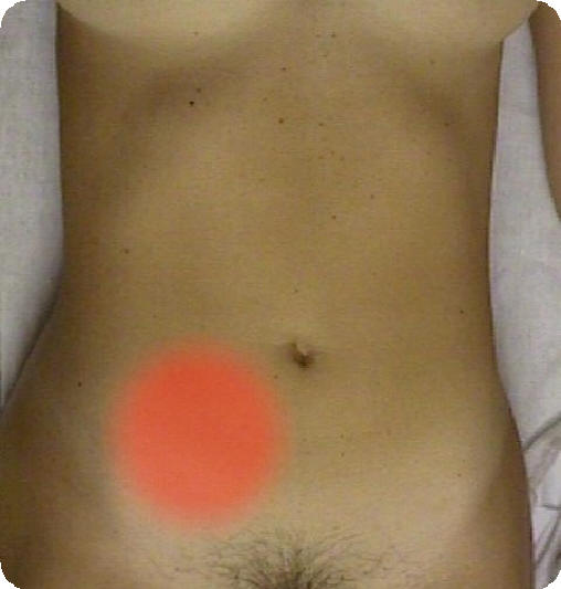
Signs of peritoneal irritation in acute appendicitis
-
Progression of pain
-
Begins in umbilical area
-
Localizes in right lower quandrant
-
Guarding/muscular rigidity
-
Voluntary guarding by tightness of muscle against palpation.
-
b. Involuntary resistance, progressive abdominal rigidity. Patient is unable to relax muscles. Body’s protective function against pain.
-
Localized tenderness - usually in RLQ or right flank pain.
-
Rectal exam reveals right sided rectal tenderness. May indicate inflammatory process other than appendicitis.
-
Rebound tenderness
-
Rovsing’s sign (referred tenderness): tenderness/pain in RLQ during left sided pressure.
-
Referred rebound tenderness
-
Psoas sign: An increase in pain from passive extension of the right hip joint that stretches the iliopsoas muscle (Tabers Medical Dictionary, 1989).
-
Place right hand above right knee of the patient.
-
Have the patient flex right knee against resistance.
-
Alternatively, have the patient turn to side, extend right leg at right hip.
-
Pain with maneuvers suggests irritation of Psoas muscle.
-
Obturator sign
-
Flex patients right thigh at hip with right knee bent.
-
Internally rotate the leg at the hip.
-
Pain elicited suggest irritation of obturator muscle.
-
Cutaneous Hyperesthesia: Increased sensitivity to sensory stimuli, such as pain or touch.
-
At a series of points down the abdominal wall, gently pick up skin folds between finger and thumb without pinching the skin.
-
Localized pain elicited in the RLQ may accompany appendicitis.
-
Acute Cholecystitis: Inflammation of the gallbladder.
-
RUQ pain and tenderness
-
Murphy’s sign: When the inflamed gallbladder is palpated by pressing the fingers under the rib cage, deep inspiration causes pain because the gallbladder is forced down to touch the fingers.
-
Hook fingers under costal margins on the right.
-
Have the patient take deep breath.
-
Sharp increase in tenderness with sudden stop in inspiration is positive.
-
Positive sign is indicative of gall bladder disease.
-
Intra-abdominal mass vs. abdominal wall mass
-
Have the patient tighten abdominal muscles wall.
-
Mass in abdominal wall remains palpable where as intra-abdominal mass will be obscured.
|
|
Approved for public release;
Distribution is unlimited.
The listing of any non-Federal product in this CD is not an endorsement of the
product itself, but simply an acknowledgement of the source.
Bureau of Medicine and Surgery
Department of the Navy
2300 E Street NW
Washington, D.C
20372-5300 |
Operational Medicine
Health Care in Military Settings
CAPT Michael John Hughey, MC, USNR
NAVMED P-5139
January 1, 2001 |
United States Special Operations
Command
7701 Tampa Point Blvd.
MacDill AFB, Florida
33621-5323 |
*This web version is provided by
The Brookside Associates Medical Education Division. It contains
original contents from the official US Navy NAVMED P-5139, but has been
reformatted for web access and includes advertising and links that were not
present in the original version. This web version has not been approved by the
Department of the Navy or the Department of Defense. The presence of any
advertising on these pages does not constitute an endorsement of that product or
service by either the US Department of Defense or the Brookside Associates. The
Brookside Associates is a private organization, not affiliated with the United
States Department of Defense.
Contact Us · · Other
Brookside Products

|
|
Operational Medicine 2001
Contents
|
|
 |
|
FMST Student Manual Multimedia CD
30 Operational Medicine Textbooks/Manuals
30 Operational Medicine Videos
"Just in Time" Initial and Refresher Training
Durable Field-Deployable Storage Case |
|