Hospital Corpsman Sickcall Screener's Handbook
BUMEDINST 6550:9A
Naval Hospital Great Lakes
1999
Dermatology Disorders and Examination
Allotted time:
References:
Terminal learning objectives: Given a simulated patient with simulated dermatological symptoms, the student will be able to recognize potential problems and properly perform the needed exam.
Enabling learning objectives:
The student will be able to identify different types of lesions.
The student will be able to identify the different types of common dermatological conditions.
The student will be able to identify the signs and symptoms of common dermatological conditions.
-
The student will be able to identify the treatment of common dermatological conditions.
-
The student will be able to identify the different components of the dermatological exam.
-
The student will be able to identify the variances of skin color.
-
Techniques of exam
-
Inspect and palpate for:
-
vascularity, evidence of bleeding, or bruising
-
color
-
moisture, dryness, sweating, oiliness
-
use back of fingers to check temperature
-
texture
-
thickness
-
mobility and turgor
-
Observe any lesions of the skin for:
-
location and distribution
-
grouping and arrangement
-
types of lesions
-
note color of lesions
-
Inspect and palpate
-
nail beds of fingers and toes
-
the hair for quantity, distribution, texture
-
Common dermatological conditions
-
Contact dermatitis: a chronic or acute inflammation produced by substances coming into contact with the skin. Classic examples are poison ivy/poison oak.
-
Signs/symptoms
-
itching
-
scaling
-
rash
-
redness or swelling
-
generally discrete areas are affected, i.e. only those that were in contact with irritant.
-
Treatment
-
determine/eliminate causative agent
-
keep area clean and dry
-
antibiotics (if infection has developed)
-
hydrocortisone cream (HC) 1% TAM 0.1% BID on affected area
-
refer to MO for severe or extensive cases (i.e. prednisone TX)
-
Acne: common inflammatory pilosebaceous disease characterized by comedones, papules, pustules, inflamed nodules, and pus (purulent) filled cyst.
-
Types
-
comedones: 2 types
-
open: black heads
-
closed: white heads
-
Signs/symptoms
-
Inflamed pustules
-
Superficial cysts and pustules
-
Commonly on face, neck, chest, back, and shoulders
-
Treatment
-
Wash face with mild soap with warm water (recommend Dove soap)
-
5-10% benzoyl peroxide applied in the morning after washing
-
T-stat pads (E-mycin 2%
topical) bid after washing. -
Retin-A cream (for dry skin) or gel (for oily skin) 0.025% applied qhr after
washing
-
Tetracycline 500mg qid or E-mycin 500mg bid (in severe or refractory cases).
-
If not responsive, consult with MO regarding Dermatology consult for Accutane
therapy
-
Urticaria (hives)
-
Signs/symptoms
-
pruritus
-
wheals
-
erythema and edema
-
angioedema - diffuse swelling of loose subcutaneous tissue.
NOTE: Edema of upper airway may produce respiratory distress.
-
Treatment
-
Remove offending agent if possible (May be difficult to detect)
-
Discontinue all non-essential meds
-
Oral antihistamine - Diphenhydramine HCL (Benadryl) 50-100mg q4h or
Atarax 25-50mg tid to qid
-
For pharyngeal or laryngeal angioedema, give Epinephrine 1:1000 0.3 ml SC and refer to MO or ER STAT
-
Herpes Simplex: (cold sores) a recurrent viral infection characterized by a sudden appearance of small vesicles on base of the skin or mucous membranes, often around the mouth. Generally Type I but can be Type II from oral-genital sexual contact.
-
Signs/symptoms
-
Tenderness, pain, mild burning at the site, headache, malaise, fever prior to eruptions. -
Itching/tingling sensation
-
Grouped vesicles
-
Typically painful
-
Factors that precipitate lesions: sunburns, food allergy, onset of menstruation, and
disease that may produce a fever
-
Treatment
-
Usually heal in 2-6 weeks
-
Use sunscreens
-
Systemic antibiotics
-
No corticosteroids
-
Drying lotions
-
Antivirals i.e. - Zovirax (Acyclovir) 200mg q4h five times a day for 5d
-
Herpes Zoster (shingles): an acute viral infection of the CNS characterized by vesicular eruptions and neuralgic pain in areas supplied by peripheral sensory nerves (dermatomes). Same virus that causes chickenpox. The pain in Herpes Zoster may resemble abdominal disease, pleurisy, MI, or migraine headaches depending on the location of involved nerve. One attack usually confers immunity. Must be seen by MO if on face
-
Signs/symptoms
-
4-5 days prior to eruption
-
Chills, fever, malaise, GI disturbances, and with or without pain along site of eruption.
-
May have regional lymphadenopathy.
-
4-5 days
-
Characteristic crops or vesicles on an erythematous base.
-
Involved zone is usually excessively sensitive to stimuli.
-
Pain may be severe.
-
Vesicles begin to dry and scab on about 5th day.
-
Generally all are crusted and falling off in 2-3 weeks.
-
Diagnosis
-
Difficult in pre-eruption stage.
-
Made readily after the vesicles appear.
-
Treatment
-
Zovirax 800mg q4h 5 times a day for 7-10 days (Must be given at onset or will not be helpful).
-
Giving ASA with/without Codeine for pain administration and
corticosteroids may relieve pain in severe cases.
-
Refer to MO.
-
Chicken pox (varicella)
-
Signs/symptoms
-
9 to 21 days after exposure and 2-3 days before lesions appear, will have mild headache, moderate fever, and malaise is present.
-
Itchy "teardrop" vesicle with red areolas.
-
Individual lesions progress from macule to papule to vesicle with in 6-8 hrs.
-
Upper trunk is most frequent site affected.
-
Starts centrally and spreads distally.
-
Spread by airborne droplets.
-
Pneumonia is the most common serious complication in adults.
-
Diagnosis
-
Rule out
-
Secondary syphilis (RPR)
-
Impetigo (C&S of lesion)
-
Infected eczema (history)
-
Insect bites (history)
-
Drug rashes (history)
-
Contact dermatitis (history)
-
Treatment
-
Zovirax 800mg qid for 5 days
-
refer to MO
-
Isolate from people who have not been previously exposed. (Will require convalescent leave if in barracks).
-
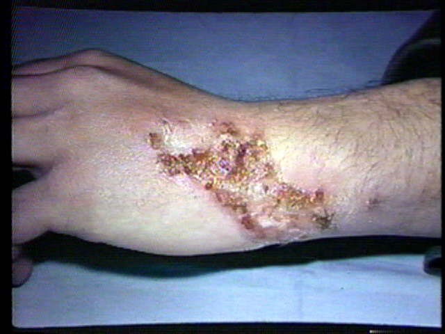
Impetigo: a superficial skin infection caused by staphylococcus or streptococcus infection
-
Signs/symptoms
-
arms, face, and legs are commonly affected areas.
-
May follow superficial trauma, break in skin, pediculosis, scabies, fungal, dermatitis, or insect bites.
-
Lesions vary in size.
-
Lesions progress rapidly from maculopapule to vesiculopustules or bullar to exudate. Lesions are often crusted and honey colored.
-
Itching.
-
Treatment:
-
Dynapen (Dicloxacillin) 250mg or Kefelex
(Cephalexin) 250mg qid for 10days
-
Tap water compresses
-
Keep area clean and dry.
-
Topical antibiotic cream
-
treat underlying cause
-
Eczema is characterized as a dermatitis commonly located to the legs, arms, and hands. Presents as dry, "cracked", fissured skin. (More common in older persons). Can be a genetic tendency for dry skin.
-
Signs/symptoms
-
Dry/cracked skin with red fissures and sometimes lichenification.
-
Pruritus (burning sensation)
-
Often a history of too frequent bathing in hot, soapy baths/showers.
-
Diffuse skin involvement without identifiable borders.
-
Distribution is generalized.
-
Itching
-
Treatment
-
Increase contact with humidified air (above 50%). Room humidifiers in the bedroom are helpful.
-
Tepid water baths with bath oils and immediate liberal application of emollient ointments.
-
HC 1% AAA qid until resolved.
-
topical applications of alpha-hydroxy acids, such as glycolic acid and lactic acid are effective.
-
Furuncles and carbuncles
-
Definition
-
Furuncles: (abcess or boil) are acute, tender perifollicular inflammatory nodules caused by staphylococci.
-
Carbuncles: a group of furuncles, often extensive, local sloughing with slow healing.
-
Location
-
Furuncle - neck, face, breast, buttocks
-
Carbuncle- neck, back or trunk, thighs
-
Treatment
-
Treat with intermittent moist heat soaks. Allow to come to head and drain. Extensive incision may spread the infection. -
For the nose or central facial area, it should be treated with systemic antibiotics. -
For multiple carbuncles and furuncles, treat same as "b"
-
Cellulitis: an acute inflammation within the soft tissue characterized by hyperemia, leukocytic infiltration and edema.
-
Signs/symptoms
-
Skin temperature is hot.
-
red and edematous
-
lymphangitis (streaking) and lymphadenopathy
-
Diagnosis depends on clinical findings -
Treatment
-
Dicloxacillin 250mg qid or a cephalosporin orally
-
Rocephin 1gm IM when first seen.
-
Rest and elevate affected part
-
Moist heat
-
Refer to MO
-
Possible admission to hospital.
-
Outline area in pen to determine progression/regression during follow up.
-
Lymphangitis: an acute inflammation of the lymphatic channels
-
Signs/symptoms
-
Red streaks, tender and irregular, develop and extend proximally.
-
Regional lymph nodes are enlarged and tender.
-
cFever, chills, tachycardia, headache, and leukocytosis
-
Diagnosis
-
Red irregular streaks, extending toward regional lymph nodes from peripheral lesion on an extremity indicates
lymphangitis.
-
Treatment
-
Refer to MO
-
Lymphadenitis: inflammation of a lymph node.
-
signs/symptoms
-
may be asymptomatic or may have pain and tenderness
-
abscess may be present
-
ask about weight loss/night sweats
-
if positive refer to MO
-
diagnosis
-
lymphadenitis and its cause is usually apparent
-
if multiple sites, refer to MO
-
treatment
-
treat underlying cause
-
hot/wet applications
-
abscesses require surgical drainage
-
RTC in 24 hrs for F/U
-
Warts (verrucae) are a common contagious, benign epithelial tumor caused by papovirus
-
signs/symptoms
-
sharply demarcated
-
rough surfaced
-
round or irregular
-
firm, light gray, yellow, brown, grayish black tumors 2-10mm in diameter.
-
appears on fingers, elbows, knees, face, scalp
-
diagnosis by appearance
-
treatment
-
refer to derm clinic or consult with MO
-
Pityriasis Rosea: a self limited, mild inflammatory skin disease characterized by scaly lesions, occurs at any age, unknown infectious agent.
-
signs/symptoms
-
herald or mother patch found on trunk 2-10cm in size
-
patch usually proceeds full rash and is usually missed
-
erythematous, rose or fawn colored
-
scaly
-
resembles ringworm
-
may itch, Christmas tree pattern
-
diagnosis
-
clinically with woods lamp (cobalt blue)
-
must be able to differentiate from the following
-
psoriasis
-
secondary syphilis
-
If unsure, refer to MO.
-
treatment
-
no specific treatment; remission occurs within 4-5 weeks
-
reassure patient
-
oral antihistamines and a topical corticosteroid
-
If patient has severe itch, may give Prednisone 10mg qid until itching subsides then decrease over a 14 day period (can also give 3-5 day burst)
-
Pediculosis: (capitis, corpus, pubis) is an infestation by lice
-
signs/symptoms/diagnosis
-
capitis
-
corpus
-
uncommon under good hygiene
-
nits found in body hair
-
body louse inhabit seams of clothing worn next to skin
-
itching
-
lesions are common on the shoulders, buttocks, and abdomen
-
pubis
-
infests over anogenital region
-
OVA are attached to skin at base of hairs
-
scattering of minute specks
-
sometimes seen as bluish spots on the skin
-
treatment
-
wash and dry affected areas
-
1% gamma benzene hexachloride shampoo
(kwell) apply to affected areas. Apply only to dry hair, and work well into the affected areas. Leave on for 4 minutes. Apply some water and work into lather. Rinse all lather away.
-
reevaluate in 7 days
-
dead nits must be combed from hair
-
decontaminate combs, clothing, bedding, etc by washing at 140F
-
Scabies: a parasitic skin infection characterized by superficial burrows, intense pruritis and secondary inflammation seen as fine wavy dark lines.
-
signs/symptoms/diagnosis
-
pruritis marked, intense at bedtime
-
lesions are the burrows
-
lesions occur predominantly on the following
-
burrows may be hard to find due to scratching and/or secondary lesions
-
treatment
-
Kwell lotion or cream applied from tip of chin to tip of toes. Leave on 12 hours and wash off. Reevaluate in 12 weeks. -
10% Crotamiton
(Eurax) generally given to young children or pregnant patients. Apply to whole body from chin down. Repeat in 24 hrs. Wash off in 48 hours after last application.
-
Tineas (superficial fungal infections)
-
signs/symptoms
-
Capitis (head)
-
Cruris (jockitch)
-
Pedis (athletes foot): usually affects 4th and 5th toe spreading to plantar area. Lesions appear as macerated areas with scaling borders. -
Corporis (ring worm): lesions with borders spread peripherally and clear centrally. Typical scaly borders
-
Diagnosis confirmed by KOH or culture.
-
Treatment
-
antifungal creams/lotions for 3-4 weeks
-
Refer to MO in severe cases for possible Griseofulvin or
Ketoconazole therapy.
-
Tinea capitis does not respond well to topical treatment.
-
Tinea versicolor: an infection characterized by multiple usually asymptomatic patches of lesions varying in color from white to brown.
-
signs/symptoms/diagnosis
-
tan, brown, white, slightly scaling lesions seen on neck, chest, abdomen
-
areas do not tan
-
wood light exam
-
treatment
-
Selenium Sulfide
(Selsun shampoo): use for one week at bed time like a lotion, then wash off in AM. Continue weekly applications afterwards, applying in shower and washing off after 10 minutes.
-
Watch for skin irritation
-
Advise patient that recurrence is likely and it doesn’t need to be treated unless patient desires it.
-
PFB (pseudo folliculitis barbae)
-
signs/symptoms/diagnosis
-
Ingrown hairs resulting in papules, usually on upper neck.
-
Treat in accordance with Navy or USMC PFB program.
-
Retin-A at bedtime, Vioform HC in AM, & Benzoyl peroxide 5% in the AM.
-
Dyshydrosis (pompholyx)
-
signs & symptoms
-
Deep seated itchy vesicles on palms, sides of fingers and soles. Unkown etiology.
-
Treatment
-
Topical corticosteroid cream tid
-
cold wet compress
-
oral E-mycin, refer to MO
-
sunburn (acute)
-
Signs/symptoms
-
appears 1 to 24 hours and will usually pass its peak in 72 hours
-
skin changes range from
-
erythema with scaling
-
pain
-
swelling tenderness
-
blisters
-
fever, chill, shock may appear if a large portion of body surface is affected
-
secondary infection is the primary complication.
-
Treatment
-
initially avoid oils and creams
-
Tylenol or Aspirin for pain relief.
-
Photosensitivity: skin eruptions in response to exposure to sunlight.
-
signs/symptoms
-
erythema/dermatitis
-
urticaria
-
erythema multiform like lesions
-
bullae
-
chronic thick scaling patches
-
causes
-
numerous factors (many unknown)
-
SLE or cutaneous LE
-
herpes simplex
-
drugs such as TCN and Vibramycin
-
treatment
-
avoidance of sunlight
-
wear protective clothing
-
sun screen
-
R/O other factors
-
refer to MO
-
Scarlatina (scarlet fever)
-
Signs/symptoms
-
sore throat
-
chills, fever
-
strawberry tongue
-
cervical lymphadenopathy
-
rapid pulse
-
rash on abdomen, chest
-
a sequela to a streptococcal infection
-
Treatment: Penicillin V 250mg qid for 10 days
(E-mycin 250mg qid for 10 d)
-
Anatomy and physiology
-
Examination
-
Whole body: look at whole body. Compare one area to another. Look at the body as a whole not just the affected areas. -
Skin layers:
-
epidermis - thin outer layer that acts as a barrier -
dermis - lies just below epidermis. Serves 3 major functions
-
protects body from trauma
-
contains sensory nerve endings
-
contains sebaceous glands
-
subcutaneous - lies below dermis and acts as an insulator for body and is the main depository of fat
-
Inspection: Look at the area affected in comparison with the rest of the body. Note color, texture, temperature and deformities. A culture and sensitivity (C&S) or Potassium Hydroxide (KOH) test could assist in your diagnosis of localized lesions.
-
|
 |
|
Military
Medical CDs
Textbooks, Manuals, Instructions,
Videos
On-line Resources
www.brooksidepress.org |
Techniques of exams
-
skin - inspect and palpate
-
color
-
normal skin pigment greatly varies from person to person and is best determined by a part to part comparison. -
color changes
-
erythema: a reddish tint due to increased blood flow or RBC’s seen in sunburn and high fevers. -
cyanosis: a bluish tint brought on by a lack of oxygenated blood. Seen in pneumonia or congenital diseases, due to shunting of blood from the right to the left side of the heart. -
pallor: a whitish tint brought on by a lack of color due to a decrease in hemoglobin content. -
greenish-yellow: due to an increased amount of bilirubin content in the skin of sclera, more well known as jaundice. -
orange-yellow: a pigment brought on by an increased amount of carotenoid in the skin and unlike jaundice will not be noted in sclera. Usually caused by ingestion of excess amounts of food with carotene. -
gray: a gray color may be noted due to a deposition of mineral salts such as gold, silver, or bismuth brought on by overuse of
silvadene or pepto-bismal. -
increased/decreased pigmentation: a darkening or lightening of the skin brought on by excess or absence of melanin in the body. -
localization pigmentation: pigmentation from the injection of foreign substances.
-
moisture: dryness, sweating or oiliness
-
temperate: generalized warmth with fever, coolness in hypothyroidism, local warmth with inflammation.
-
texture: roughness or smoothness
-
mobility and turgor: lift a fold of skin and note it’s ease in movement and the speed it returns
-
lesions
-
The principle is to accurately describe the skin lesion, which should include:
-
distribution and location
-
grouping and arrangement
-
eruptions consist of one or more lesions which can be either discrete or confluent -
Certain lesions effect only exposed areas of the body such as poison ivy and others prefer specific locations such as herpes zoster (which occur only in dermatonal patterns).
-
contour: describe the shape as best as possible
-
consistency: note whether the lesions are basically the same size, contour, color, etc.
-
size
-
Types of skin lesions
-
Primary lesions: circumscribed, flat, non palpable
-
macule - small, up to 1cm freckle or petechia
-
patch - >1cm, vitiligo
-
Palpable, elevated solid mass
-
papule - up to 1/2 cm, elevated nevus
-
plaque - flat, elevated surface > 1/2cm often formed by coalescence of papules
-
nodule - 0.5 to 2cm deeper and firmer than papule
-
tumor - >2cm
-
wheal - somewhat irregular, transient, superficial area of localized skin edema. Insect bites and hives.
-
-
Circumscribed superficial elevations of the skin
-
Vesicle: up to 1/2cm filled with serous fluid i.e.. herpes simplex
-
Bulla: >1/2cm filled with serous fluid, 2nd degree burn, blisters
-
Pustules: filled with pus (purelence). Acne, impetigo
-
Secondary lesions - results from changes in primary lesion
-
loss of skin surface
-
erosion: Loss of superficial epidermis. Moist but does not bleed. Old chicken pox lesions. -
ulcer: Deeper loss of skin surface involving epidermis and dermis. May bleed and scar. -
fissure: Linear crack in skin involving epidermis and dermis. Athletes foot
-
Material on skin surface
-
crust: Dried residue of serum, pus, blood. -
scale: Thin flake of exfoliated epidermis.
-
Miscellaneous lesions
-
Lichenification: thickening and roughening of epidermis with increased visability of normal skin furrows.
-
Excoriation: abrasions or scratch marks
-
Keloid: hypertrophic scars
-
Vascular lesions: unduly dilated superficial veins
-
Telangiectasia: localized fine red lines due to dilated blood vessels which may be capillaries or arterioles.
-
Spider angiomas: Cutaneous lesions found in areas drained by the superior vena cava and is characterized by a central, red, pulsating vessels with fine, small vessels which radiate out like legs of a spider over a reddened area of about 10mm in diameter.
-
Bleeding lesions: (purpura) Which strictly means a disorder characterized by a hemorrhage into skin.
-
petechine: tiny red or brown capillary hemorrhage not more than 0.5mm in diameter which is located within the skin papillae. -
ecchymosis: (or bruises) This is a larger hemorrhage which can range from several millimeters to several centimeters.
-
An important test of your examination capabilities is the ability to distinguish the difference between pre-malignant and malignant lesions.
-
pigment nevi (moles): raised, dark brown or black lesions. May contain hair, vary in diameter from 1mm to 10mm
-
common, usually benign, may develop into malignant melanomas
-
Suspicious characteristics include:
-
assymetry
-
border irregularity
-
color changes
-
diameter
-
Accurate history is needed & should include:
-
length of time
-
changes in characteristics
-
any evidence of rapid changes in size, color, or consistency. Refer to MO ASAP.
-
Hair: Production and loss has many normal variations.
-
Baldness: unexpected hair loss, not uncommon in the aged or middle aged. Is hereditary. A sudden unexpected loss of hair may be a sign of underlying problems.
-
A change in hair color or texture, such as thinning, fine, silky hair is often associated with hypothyroidism. Dry, brittle hair which disappears from lateral portions of the eyebrow are associated with hypothyroidism, and medial portions with leprosy.
-
Excessive amount of hair growth in women, or "hirsutism" is highly uncommon. Could be due to a tumor or endocrine disorder.
-
Nails: finger and toenails grow at approximately 1mm every 10 days and usually are the first place that cyanosis is seen. Bitten or mutilated nails may indicate an emotional or personality disorder.
-
Clubbing
-
Could be a normal family trait.
-
Characterized by an extension of the horny layers of the skin over the nail.
-
Proximal portion of the wall is elevated eliminating the angle between the nail and eponychium. Soft upon palpation of nail bed.
-
As with most disorders, the more severe the clubbing, the more evident it will be.
-
Diagnostic of congenital heart disease, chronic pulmonary disease or arteriovenous shunts.
-
Inspect nails for chronic infections such as:
-
Splinter hemorrhages - a thin, brownish flame shaped line(s) in the nail beds which could be a sign or start of a serious infection, common in a subacute bacterial
endocarditis.
-
Examination of masses: A swelling or tumor which is larger than two centimeters in diameter.
-
When describing a mass, make sure that its done accurately. Use the list below for your description:
-
location
-
shape
-
depth
-
consistency
-
mobility
-
tenderness
-
temperature
-
color of overlying skin -
Lymph nodes: a special type of mass. They are not normally palpable due to size (1-5mm diameter) with softness and mobility. They may become inflamed, causing their size to dramatically increase. Most common cause of enlargement is due to infection within the body from which lymphatic channels drain toward the node.
-
Problems occur when metastases from neoplasms get trapped into the node. This leads to enlargment and transfer of disease from one part of the body to another. Shows as systemic disorder such as:
-
Infections
-
ubella
-
mono
-
HIV
-
lymphoid tissue disease
-
Hodgkin’s disease
-
lymphomas
-
leukemia’s
-
Lymph nodes may enlarge from several different causes. With each cause they will feel slightly different.
-
Guidelines for basic consistency:
-
Moderate sized node which is firm, seperate and tender, denotes a node which is draining infection.
-
Stony hard seen in metastatic diseases.
-
Lymphatic neoplasm are often firm or rubbery
-
Three areas which the nodes are easily palpable:
-
neck
-
axilla
-
inguinal
-
When lymphadenopathy is found or noted, an accurate description and exact location is very important. Any lymphadenopathy without obvious cause must be referred to an MO.
|
|
Approved for public release;
Distribution is unlimited.
The listing of any non-Federal product in this CD is not an endorsement of the
product itself, but simply an acknowledgement of the source.
Bureau of Medicine and Surgery
Department of the Navy
2300 E Street NW
Washington, D.C
20372-5300 |
Operational Medicine
Health Care in Military Settings
CAPT Michael John Hughey, MC, USNR
NAVMED P-5139
January 1, 2001 |
United States Special Operations
Command
7701 Tampa Point Blvd.
MacDill AFB, Florida
33621-5323 |
*This web version is provided by
The Brookside Associates Medical Education Division. It contains
original contents from the official US Navy NAVMED P-5139, but has been
reformatted for web access and includes advertising and links that were not
present in the original version. This web version has not been approved by the
Department of the Navy or the Department of Defense. The presence of any
advertising on these pages does not constitute an endorsement of that product or
service by either the US Department of Defense or the Brookside Associates. The
Brookside Associates is a private organization, not affiliated with the United
States Department of Defense.
Contact Us · · Other
Brookside Products

|
|
Operational Medicine 2001
Contents
|
|
 |
|
FMST Student Manual Multimedia CD
30 Operational Medicine Textbooks/Manuals
30 Operational Medicine Videos
"Just in Time" Initial and Refresher Training
Durable Field-Deployable Storage Case |
|