|
Examining the Heart
|
![]()
An electric signal starts at the pacemaker and
spreads through the heart. |
![]()
Blood collects in the right atrium and is pumped
into the right ventricle, then on to the lungs. Blood returns from the lungs to
the left atrium, is pumped to the left ventricle, and then out through the
aorta. |
The heart is located directly behind the breast
bone (sternum) and slightly to the left.
 Palpation
(feeling) Palpation
(feeling)
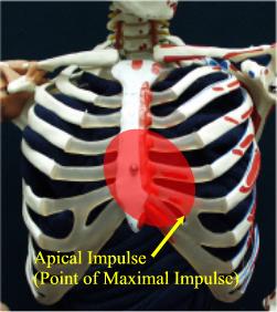
Use the palm of your hand to feel the chest wall for the "Point of
Maximal Impulse" (PMI), which is usually found at the apex of the heart.
This apical pulse is generally located in the 5th intercostal space, about
7-9 cm (the width of your palm) to the left of the midline.
In a noisy place, feeling for the apical pulse
may be the most examination of the heart that is possible.
The apical pulse should be regular, with a
rhythmic tap, tap, tap. If you cannot easily feel it, have the patient
roll slightly to the left, bringing the heart more into contact with the
chest wall.
-
The apical pulse should always be closer to
the midline than the mid-clavicular line.
-
If lateral to the MCL, this suggests cardiac
enlargement.
-
In patients with pericardial effusions, you
may not be able to locate the apical pulse.
-
If you feel a vibration or buzzing sensation
while feeling the apical pulse, this suggests a heart murmur. This
finding is known as a "thrill."
-
"Thrills" or murmurs from other areas of the
heart are best felt through the bones of the chest wall.
|
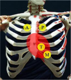
Listen to the heart sounds from each of the
4 heart valves at these locations. |
Auscultation (listening)
In a quiet place, use a stethoscope to listen to the heart sounds.
Listening to the heart can be helped by having the patient sit up and lean
forward, or having the patient roll to the left side.
The diaphragm of the stethoscope (flat portion)
is better for hearing higher-pitched sounds. The bell (curved portion) is
better for hearing lower-pitched sounds. Press the stethoscope lightly
against the skin, just enough to form an air seal around the edges. Have
the patient exhale and then stop breathing for a moment while you listen.
Each of the four heart valves can best be heard
at specific anatomic locations on the chest wall. While there may be some
variation from patient to patient, listen to the heart beat at each of
these areas:
-
Aortic (2nd right interspace)
-
Pulmonic (2nd left interspace)
-
Tricuspid (Lower left sternal border)
-
Mitral (Apex)
If you are connected to the Internet, you can hear actual heart sounds,
normal and abnormal, at this website.
|
 |
|
This is a mid-systolic, crescendo-decrescendo
type murmur.
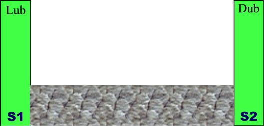
This is a pan-systolic type murmur.
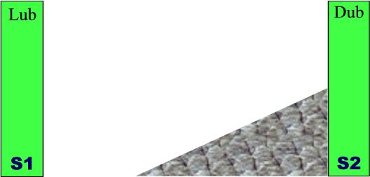
This is a crescendo, late systolic type murmur.
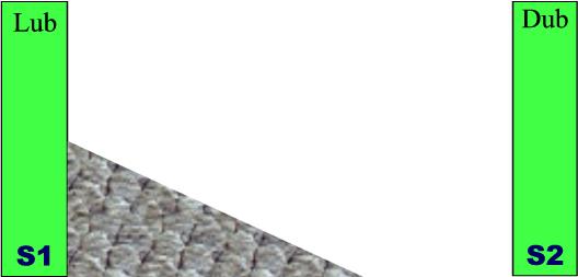
This is a decrescendo, early systolic type murmur.
|
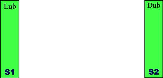
Normal Heart Sounds
Heart sounds are more complicated than a simple "lub-dub." The
first heart sound (the "lub") is called "S1." It is normally a single
sound, but may be split into two distinct sounds, like a "da-dum." While a
split S1 may be associated with various heart abnormalities, some people
have a normally split S1, that fuses into a single sound when they exhale.
The second heart sound (S2), the "dub" part of "lub-dub"
is also a single sound. It, too, can be split into two distinct sounds and
that may reflect underlying heart defects, or may be normal.
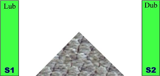
Heart Murmurs
Normally, you will not hear any heart murmurs. Murmurs are soft,
humming or buzzing sounds, occurring between the "lub" and the "dub."
(between S1 and S2). These are called "systolic" murmurs, occurring during
cardiac systole (contraction of the heart). There are also "diastolic"
murmurs, occurring after one "lub-dub" and before the next "lub-dub."
Murmurs are caused by turbulence in the blood
flow through the heart, typically as it passes through a heart valve. For
example, a damaged aortic valve might not open wide enough (aortic
stenosis) to allow blood to flow freely through it from the heart into the
aorta during cardiac systole. The turbulent flow across the stenotic valve
causes a buzzing sound, called a murmur, between the "lub" and the "dub."
The presence of a heart murmur is occasionally
normal (such as increased flow murmurs during pregnancy), but usually
indicates a cardiac abnormality.
Other Heart Sounds
Other heart sounds may be heard, the most common of which is the
"ejection click," associated with mitral valve prolapse.
|