Examining the Eyes
Atlas of Eye
Problems The eye is a hollow ball, or globe, which consists of various tissues that
perform specific functions. The globe, or eyeball, is composed of three layers.
Outer Layer - The outer layer of the eye is called the sclera. It is
the tough, fibrous, protective portion of the globe, commonly called the white of the eye.
Anteriorly, the outer layer is transparent and is called the cornea, or the window of the
eye. It permits light to enter the globe. The exposed sclera is covered with a mucous
membrane, the conjunctiva, which is a continuation of the inner lining of the eyelids. The
lacrimal gland produces tears that constantly wash the front part of the eye and the
conjunctiva. The tear gland secretions that do not evaporate flow toward the inner angle
of the eye where they drain down ducts into the nose.
|
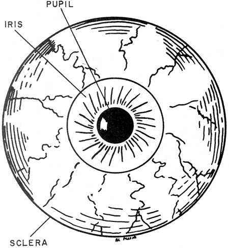
Anterior View of the Eye
Look Up
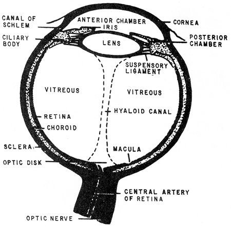
Cross Section of the Eye
The Vision Process
Ophthalmoscope View of the Eye
Normal Fundus
Look Down
Look Left and Right
|
Middle Layer - The middle layer of the eye is called the choroid. It is
a highly vascular, pigmented tissue that provides nourishment to the inner structures.
Continuous with the choroid is the ciliary body, whose muscular structure attaches to the
lens by means of suspensory ligaments and produces changes in the thickness of the lens.
This permits the eye to focus to longrange or close-up vision.
The iris is continuous with the ciliary body. It is a circular, pigmented
muscular structure that gives color to the eye. The opening in the iris is called the
pupil. The amount of light entering the pupil is
regulated through the constriction of radial/circular muscles in the iris. When strong
light is flashed into the eye, the circular muscle fibers of the iris contract, reducing
the size of the pupil. If the light is dim, the pupil dilates to allow as much of the
light in as possible. The size and reaction of the pupils of the eyes are an important
diagnostic tool.
The lens is a transparent, biconvex structure suspended directly behind the
iris. It separates the interior eye into anterior and posterior cavities. The anterior
cavity contains a watery solution alled aqueous humor, which helps to give the cornea its
curved shape. The optic globe posterior to the lens is filled with a jellylike substance
called vitreous humor, which helps to maintain the shape of the eyeball and prevents
misshaping by maintaining intraocular pressure.
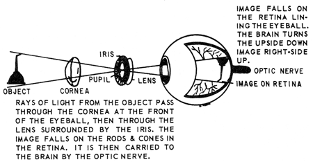
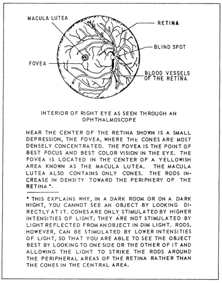
Inner Layer - The inner layer of the eye is called the retina. It contains different layers of nerve cells, rods, and
cones that are the receptors of the sense of vision. The retina is continuous with the
optic nerve, which enters the back of the globe and carries visual impulses received by
the rods and cones to the brain. The area where the optic nerve enters the eyeball
contains no rods and cones and is called the blind spot.
The rods respond to low intensities of light and are responsible for night
vision. They are located in all areas of the retina, except in the small depression called
the fovea centralis, where light entering the eye is focused, and which has the clearest
vision.
The cones require higher light intensities for stimulation and are most
densely concentrated in the fovea centralis. The cones are responsible for daytime vision.
Vision Process
Deflection or bending of light rays results when light passes through
substances of varying densities in the eye (cornea, aqueous humor, crystalline lens, and
vitreous humor). The deflection is referred to as
refraction. Accommodation is the process performed by the lens that increases or decreases
its curvature to refract light rays into focus on the fovea.
The constriction of the pupil by the iris regulates the amount of light
entering the eye. This process protects the retina from excessive stimulation and prevents
a scattering of light rays that would produce blurred vision.
A movement of the globes toward the midline, which causes a viewed object to
come into focus on corresponding points of the two retinas, is called convergence. This
gives clear, three dimensional vision.
The end receptors or nerve endings in the rods and cones that have been
stimulated by light conduct impulses to the occipital lobes of the cerebrum, where they
are interpreted into vision.
Physical exam:
-
Test visual acuity —Snellen chart at 20 feet is the best screening
method.
-
"Cover one eye and read the smallest line
possible".
-
Visual acuity is expressed as two number 20/30.
-
The first number is the distance in feet from chart, the second
the distance at which a normal eye can read the line of
letters.
-
Vision of 20/200 means that the patient can read print at 20
feet that a person with normal vision could read at 200
feet.
-
You can test visual acuity with any available print.
-
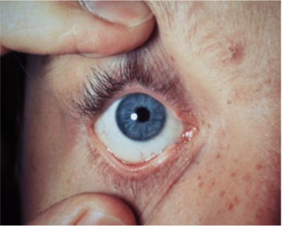
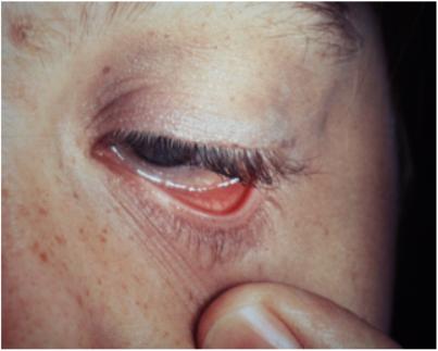
Inspection of eyelids, conjunctiva and sclera:
-
Observe eyelids for redness, swelling, and
lesion’s.
-
Inflammation of an eyelash follicle with a lump, called a sty or
hordeolum, is usually caused by staph.
-
Check the position of the upper lid — it should cover the top
part of the iris only but not the pupil.
-
Ptosis is present when the upper eyelid droops over the pupil.
-
Check the conjunctiva and sclera for redness color or discharge.
A yellow sclera indicates jaundice.
-
Ask the patient to look up as you depress both lower lids with
your thumb exposing the sclera and conjunctiva.
-
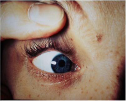
A special exam is done if you suspect a foreign body —
eversion of the upper eyelid. Ask the patient to look down, pull
downward and forward on the eyelashes. Place a "Q" tip 1
cm above the lid margin and push down on the upper lid everting
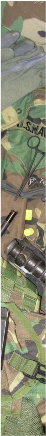
it. Alternatively, a bent paperclip can be used.
-
Pupils — Inspect the size and equality of pupils.
Test the pupillary response to light — shine light obliquely into
each eye. Look for:
-
Direct reaction (constriction of the same eye)
-
The consensual reaction (pupillary contraction in the opposite
eye).
-
Extra ocular Eye Muscles:
Ask patient to watch your finger as you move it in six directions
(think of a capital H) Watch for Nystagmus — the involuntary
rhythmic rapid movement of the eye.
Everting the lids:
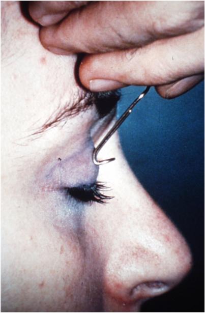
Step 1: Place the paperclip over the eyelid |
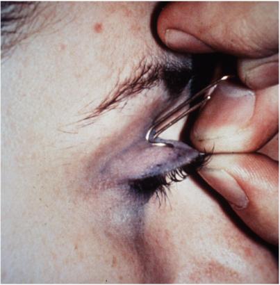
Step 2: Grasp the eyelashes and pull the eyelid outward |
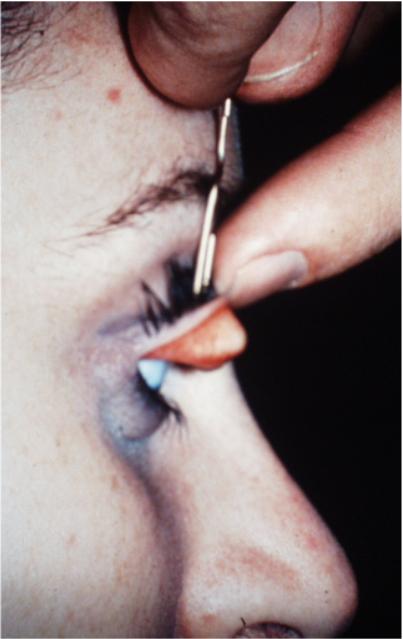
Step 3: Flip the eyelid up and over the paperclip. |
For further reading, see:
|