|
Intrauterine Device (IUD)
IUDs have been known and used for thousands of years in large domestic animals,
but only recently have they been used by humans.
Modern IUDs are easily inserted, have a very high effectiveness rate (98-99%), and are
well-tolerated by most of the women who use them. Their effectiveness continues for
varying lengths of time, depending on the type of IUD. The "Copper T 380A," used
frequently in the United States, can remain in place for 10 years before removal is
recommended.
IUDs tend to make menstrual flows somewhat heavier, crampier and longer, a
consideration in assessing the appropriateness of an IUD for any individual patient.
The Dalkon Shield
While many IUDs were known to be safe and effective, one in particular, the Dalkon
Shield, seemed to have more than its' share of problems, the most important of which was
infection. Pelvic infections, infrequent and usually minor with the other IUDs, tended to
be more frequent and more severe among Dalkon Shield users. Many of these infections were
so serious as to render the patient permanently sterile or to necessitate a hysterectomy.
There were two reasons for these infections; a design flaw and a marketing flaw. The
design flaw was located in the "tail" or string used to remove the IUD. After
insertion, the string is left protruding through the cervix so it is visible on pelvic
exam. This confirms that the IUD is correctly placed and facilitates removal at a later
date. The Dalkon Shield string was made up of many tiny plastic filaments and encased in a
plastic sheath. This design inadvertently caused the string to act as a wick, constantly
drawing vaginal bacteria up through the cervix and into the uterine cavity where they
could cause infection. The other IUDs had monofilament strings which did not have the same
wicking capacity. The design, in retrospect, predisposed the Dalkon Shield to infections.
The marketing flaw was to promote IUD among young, single women without children. These
women tended to have greater risk of exposure to sexually transmitted disease and multiple
sexual partners. They tended to be more likely to seek medical attention late in the
course of the illness. The consequences of permanent infertility among these young women
was devastating.
While the design and marketing flaws of the Dalkon Shield are of primarily historical
interest, the lessons learned at a terrible cost should not be forgotten in looking at
more modern IUDs.
Infection
With the newer designs, the risk of infection has been significantly reduced. Sooner or
later, about 3-5% of IUDs will be removed because of infection. Most of these infections
are minor, with mild symptoms of vague pelvic discomfort, painful intercourse and possibly
a low-grade fever. The uterus is tender to palpation although the adnexa usually are not.
Treatment of such mild infections generally involves prompt removal of the IUD, oral broad
spectrum antibiotics and complete resolution of symptoms. Infertility following such mild
infections is uncommon.
With the less common, serious infections, a high fever can be found, movement of the
cervix causes excruciating discomfort and the adnexa are extremely tender. In addition to
prompt removal of the IUD, IV antibiotics are recommended to treat this moderate to
severe PID. In these cases, recovery is generally slow (days to weeks) and infertility is
a distinct possibility.
Perforation
The overall risk of perforation of the IUD through the
uterine wall is about 1 in 1,000. Most of these occur during the insertion of the IUD or
shortly thereafter. More common than perforation is the "disappearance" of the
IUD string. While such a disappearance may suggest the possibility of perforation, a more
likely explanation is that the string has coiled up inside the cervical canal or even
inside the uterus.
A truly perforated IUD is usually removed from the abdominal cavity
with laparoscopic or open surgery.
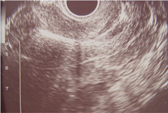
Ultrasound scan showing a Copper T IUD
positioned normally in the fundus. |
Missing IUD String
When confronted with a missing IUD string, most clinicians will gently probe the
cervical canal to see if they can tease the string back down through the os. A
cotton-tipped applicator or a Pap smear brush works well for this purpose. Once the string
is brought down into the vagina (and about 3/4 will be found this way), nothing further
needs to be done.
If the string is not inside the cervical canal, then further evaluation and treatment
will be needed from an experienced and well-equipped gynecologic consultant. X-ray can
confirm that the IUD remains somewhere within the pelvis. Ultrasound can demonstrate the
presence of the IUD inside the uterine cavity. For an IUD which is clearly inside the
uterine cavity but whose string has retracted into the cavity, a careful judgment must be
made.
In some circumstances, the IUD is removed with an IUD hook, D&C or hysteroscopy,
and a new once replaced. In other circumstances, it may be appropriate to leave the IUD
where it is until the 10 years have expired before removing it.
Pregnancy
IUDs are very effective at preventing pregnancy, but there is a small failure rate of
about 1-2% each year.
If pregnancy occurs, it is important to remove the IUD immediately (that day). The
normal spontaneous miscarriage rate is about 18-20%. For women who conceive despite an
IUD, the miscarriage rate is about 25% when the IUD is removed immediately. If the IUD is
left in place, the miscarriage rate increases to about 50%, and many of those are septic
mid-trimester losses which are particularly unpleasant and which are associated with
subsequent infertility in some cases.
If deployed, even the relatively inexperienced health care provider can remove the IUD
because: 1) it is simple and easy to do, and 2) delaying removal for several days until a
more experienced provider can see the patient risks retraction of the string up inside the
uterus, making simple removal impossible. The IUD should first be removed and then the
patient moved to a definitive care setting in anticipation of a possible miscarriage.
Ectopic Pregnancy
Should a pregnancy occur despite the presence of an IUD, there is an increased
likelihood that it will be an ectopic pregnancy. Instead of the typical rate of about 1%,
the ectopic pregnancy rate is about 5%. This means that in addition to prompt removal of
the IUD, the patient needs a careful evaluation with ultrasound and possibly adjunctive
laboratory tests to determine the presence of the pregnancy. Should an ectopic pregnancy
be found, medical and/or surgical management is usually undertaken.
In many military settings, such an evaluation may not be possible and medical
evacuation should be considered.
IUD Candidates
Contraindications to IUD use include:
-
Known or suspected pregnancy
-
Known distortion of the uterine cavity
-
PID past or current
-
Pregnancy-related infection within the last 3 months
-
Known or suspected cervical cancer
-
Undiagnosed vaginal bleeding
-
Current cervicitis or vaginitis until effectively treated
-
Wilson's disease
-
Allergy to copper
-
Impaired immune system
-
Genital actinomycosis
Insertion of the IUD
An IUD can be inserted at any time, provided the physician is confident that the
patient is not currently pregnant. Many physicians prefer to insert the IUD during a
normal menstrual flow. This provides some assurance that the patient is not currently
pregnant. Second, the cervical canal is already somewhat dilated from the menstrual flow
and so the actual IUD insertion is more comfortable for the patient. Third, there is
usually a small amount of bleeding following insertion of the IUD which will not be
noticed if the patient is currently flowing. The IUD may be inserted at the 6-week
postpartum check.
Insertion usually causes mild uterine cramping which disappears in a few minutes.
Pretreatment with a NSAID can block much of that discomfort.The use of prophylactic
antibiotics is an unresolved controversy.
Removal of the IUD
An IUD can be removed at any time, but should be removed in the presence of pelvic
infection, pregnancy, abdominal pain of uncertain cause or if the IUD is already partially
extruded. Never push a partially extruded IUD back inside the uterus as you will introduce
significant bacterial contamination into either the uterus or the abdominal cavity,
whichever area you penetrate.
After placing a vaginal speculum, visualize the cervix and the IUD string(s) protruding
through the cervical os. Grasp the strings with any convenient instrument (hemostat,
dressing forceps, ring forceps, etc.) and pull the IUD straight out with a steady, smooth,
slow pull. The IUD, by virtue of its' pliability, will fold onto itself and slide out.
Most patients will feel either no discomfort or minimal uterine cramping during removal.
They generally comment that having the IUD removed was not as uncomfortable as having it
inserted.
|