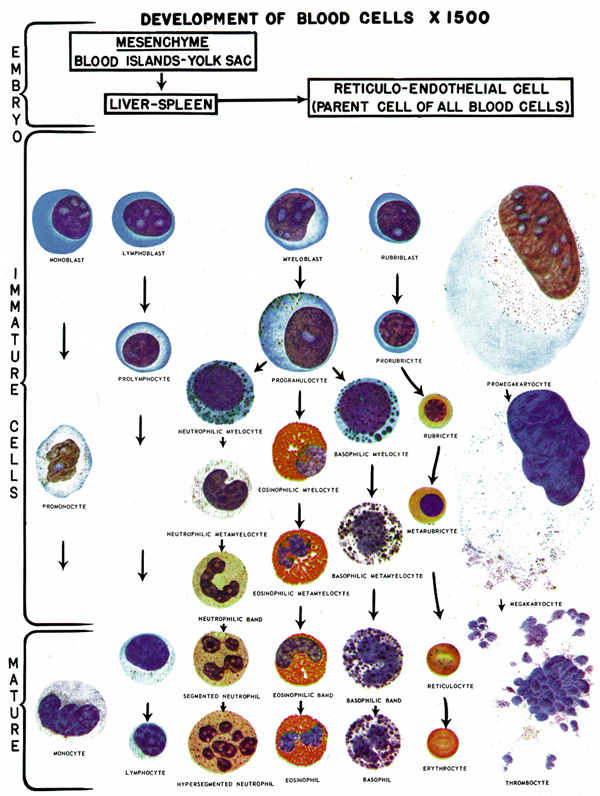
| A microscopic examination of a
stained, peripheral blood smear may be useful in evaluating blood
disorders.
Anisocytosis
- These are abnormal in size
Basophilic Stippling
- Usually seen in reticulocytes, an immature form of RBC, released in
response to strong stimulus, and seen in:
- Is nearly universally seen in cases of clinically significant lead
poisoning.
Hypochromic
- The normally pale center of the RBC is even more pale, suggesting
reduced concentrations of hemoglobin within the cell.
Malarial Stippling
- Very fine granular appearance to the RBCs, representing malarial
parasites.
- Also known as:
- Schueffner's dots
- Parasitized RBCs
Normochromic
- Normal amounts of staining for hemoglobin, suggesting normal
concentrations of hemoglobin within the cell.
Nucleated RBCs
- In an adult, RBCs are not normally nucleated.
- The presence of NRBCs indicates the body is aggressively producing
red cells and releasing them into the circulation prior to full
maturity.
- This is commonly seen in cases of significant anemia.
Poikilocytosis
- These are abnormal in shape.
- This due to improper formation of the cell membrane.
- This typically occurs in cases of severe anemia when the RBC
manufacturing system goes into overdrive, greatly increasing the
number of RBCs produced, but with lesser degrees of quality control.
Polychromasia
- Variations in staining of the RBC suggesting a more rapid production
than is usually seen.
- Commonly seen in anemias of all types, but particularly hemolytic
anemias and anemias following acute blood loss.
Reticulocytes
- An immature form of RBC, it released in
response to strong stimulus, and seen in:
Shistocytes
- Bizarre-shaped RBCs, resembling triangles or spirals.
- Associated with:
- Severe burns
- DIC
- Artificial heart valves
- Vascular spasm
Sickle Cells
- Crescent-shaped RBCs, associated with abnormal hemoglobin formation
(Hemoglobin S, SS, SC, SD or Sickle-Thallasemia.)
- Associated with hemolytic anemia.
Spherocytes
- Smaller and rounder than normal RBCs
- Associated with:
- Hereditary disease
- Post-transfusion changes
- Water dilution
Target Cells
- Smaller than normal RBCs, with a central concentration of
hemoglobin, giving it the appearance of a target.
- Associated with:
- Liver disease
- Iron deficiency anemia
- Thallesemia
|
Normal Values of the Peripheral Smear*
| Size |
Normocytic (7-8 µm) |
| Color |
Normochromic |
| Shape |
Normocyte |
| Structure |
No nucleated cells |
*These are general values taken from a variety of
sources. The actual normal values may vary from lab to lab and from one
type of testing protocol to another.
 |
Source: Operational Medicine 2001, Health
Care in Military Settings, NAVMED P-5139, May 1, 2001, Bureau
of Medicine and Surgery, Department of the Navy, 2300 E Street NW, Washington,
D.C., 20372-5300
Military Obstetrics & Gynecology
© 2003, 2004, 2005, 2006 Medical Education Division,
Brookside Associates, Ltd.
All rights reserved
|
